
|
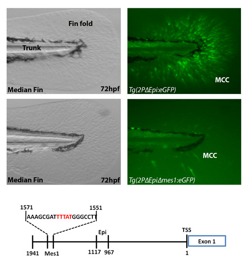
Fig. 4
Enhancer activity of 2PΔEpi drastically reduced when putative binding site for Hox (mes1 site) removed. (A-D) Zebrafish median fins at 72hpf comparing eGFP reporter expression when driven by either 2PΔEpi or 2PΔEpiΔmes1 regulatory elements. (E) Schematic of actinodin1 1941bp region of the first non-coding exon. The 2PΔEpi regulatory element is able to drive reporter expression in the migrating mesenchymal cells of the median and pectoral fin folds (Median fin: A-B, Pectoral fin: Fig. 5B). Reporter expression is drastically reduced in the median fin when mes1 site is removed from the 2PΔEpi regulatory element (C-D). No eGFP-positive cells are visible in the pectoral fin (data not shown). Mes1 site consists of a 20bp region containing the consensus Hox binding domain TTTAT (Red text) (E). 2PΔEpi regulatory contains the entire 1941bp fragment with Epi region removed (E). Two independent lines were obtained to confirmed the expression pattern of Tg(2PΔEpiΔmes1:eGFP). Brightfield (A,C) and fluorescent (B,D) images are displayed. MMC, migrating mesenchymal cells; TSS, transcription start site.