
|
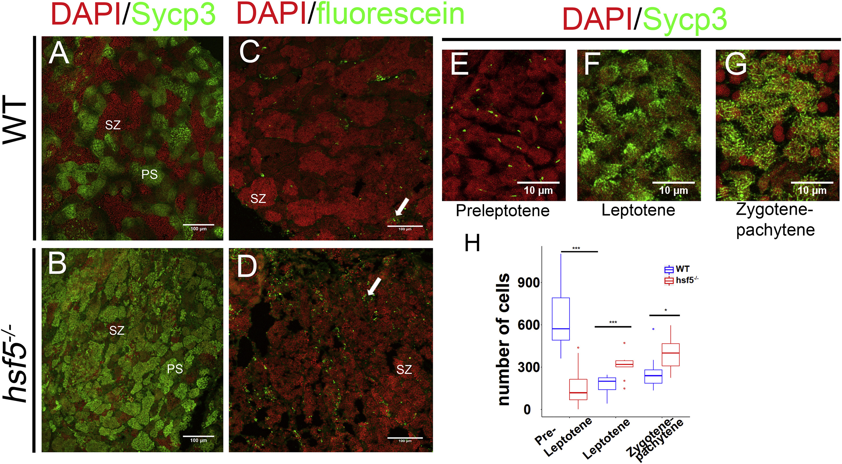
Fig. 4 hsf5−/− Testes Showed Higher Number of Primary Spermatocytes and Apoptotic Cells Compared to Wild-Type
(A and B) Representative images showing immunostaining of WT (A) and hsf5−/− testis sections (B) with anti-Sycp3 antibody. More spermatocytes were accumulated in the mutant. For (A) and (B), n = 4 biological replicates in each group. (C and D) TUNEL staining of the wild-type (C) and hsf5−/− (D) testis, showed a higher number of apoptotic cells in the latter. n = 3 biological replicates. (E–G) High-resolution confocal images of anti-Sycp3 immunostaining in WT testis showed spermatocytes at different stages of meiotic prophase-1. Sycp3 appears as small particles at one side of the cell in pre-leptotene stage (E), bouquet-shaped chromosomal arrangement in leptotene stage (F), and in reticulate manner at zygotene-pachytene stage (G). (H) Relative proportion of cells in the above three stages quantified from Sycp3-stained WT and hsf5−/− testes. Cell counts at preleptotene stage were significantly lower, whereas those at the leptotene and zygotene-pachytene stages were higher in the mutant compared to WT. For (E)–(H), n = 5 biological replicates in each group. Scale bars: 100 μm in (A)–(D) and 10 μm in (E)–(G). Difference between WT and hsf5−/− was examined with Kolmogorov-Smirnov test, ∗∗p < 0.01. The error bars indicate SD. See also Figure S5.