
|
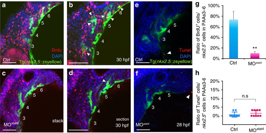
Fig. 4 Lack of stat4 inhibited proliferation of nkx2.5 PAA progenitors but not apoptosis.
(a–d) The control (a,b) and stat4 morphant (c,d) Tg(nkx2.5:ZsYellow) embryos at 30 hpf are stained by BrdU (red), 4’,6-diamidino-2-phenylindole (DAPI) (blue) as well as immunohistochemistry for ZsYellow (yellow). The single confocal sections are shown in b,d. (e,f) Projections of the pharyngeal arch region of 28 hpf control (e) and stat4 morphant (f) Tg(nkx2.5:ZsYellow) embryos are assayed for TUNEL (red) and pharyngeal arch arteries for ZsYellow (yellow). (g) Ratios of BrdU+ cells/the nkx2.5+ cells in PAAs 3–6 region across three experimental replicates (n=6 embryos/replicate in each group); Error bars indicate the s.d. Kruskal–Wallis test, **P=0.0036. (h) Ratios of TUNEL+ cells/the nkx2.5+ cells in PAAs 3–6 region, n=9 per each group, n.s.: P>0.05. White arrowheads indicate BrdU+ or TUNEL+ cells. Scale bars, 50 μm.