
|
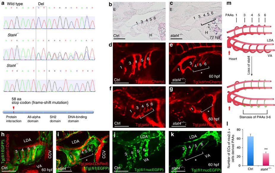
Fig. 2 Loss of stat4 leads to stenosis of PAAs 3–6.
(a) Genomic sequences of wild-type zebrafish, stat4 heterozygous mutants and homozygous mutants. The stat4 mutant has a two-base pair deletion, which causes a frame-shift mutation and a premature stop codon to form a truncated protein with 58 amino acids. (b,c) Pathological sections (10 μm per section) of wild-type and mutant embryos with Haematoxylin-Eosin staining at 72 hpf (n=12). (d,e) Confocal images of PAAs in the control Tg(kdrl:mCherry) embryos and the stat4−/−;Tg(kdrl:mCherry) line (n=9). (f,g) Images of PAAs 3–6 vessels trafficking blood flow in the control and stat4−/−;Tg(gata1:DsRed) embryos (n=9). (h,i) Images of PAAs trafficking blood flow in the control and the stat4−/−;Tg(fli1:EGFP);Tg(gata1:DsRed) line (n=8). (j–l) Control and the stat4−/−;Tg(fli1:nucEGFP) embryos show PAA endothelial cells (green) with cell number counts at 60 hpf (n=9). Error bars indicate s.d., unpaired two-tailed Student’s t-test, **P=0.003. (m) The model of PAAs 3–6 phenotype caused by stat4 deficiency is illustrated in the cartoon. Scale bars, 50 μm. LDA, lateral dorsal aorta; VA, ventral aorta; E, eye; H, heart. The brackets with asterisks highlight the malformed individual PAAs.