
|
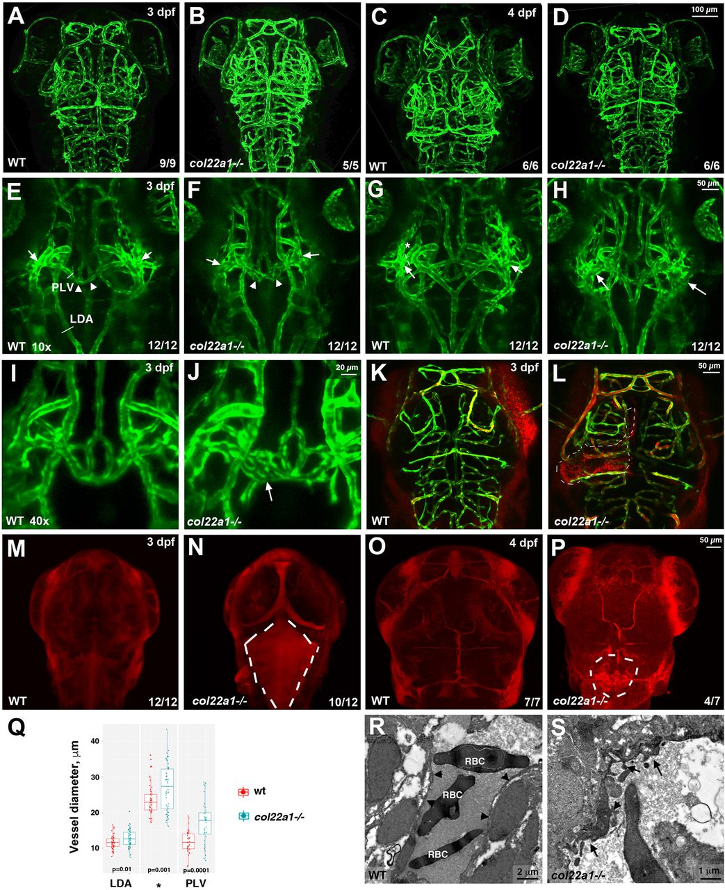
Fig. 5 Homozygous col22a1−/− mutant embryos show dilated and dysmorphic cranial vessels and have increased vascular permeability. (A-D) Vascular patterning is not affected in col22a1 mutants at 3 dpf and 4 dpf. Confocal maximal-intensity projections of live kdrl:GFP embryos, dorsal view, anterior is to the top. (E-J) Dilated and dysmorphic vessels are observed in col22a1 mutants. Note the dilated and fused vessels at the vascular branch point in the periocular region (arrows) and the dilated and dysmorphic palatocerebral vein (PLV, arrowheads). Higher magnification (40×) images of the PLV are shown in I and J. Note that a portion of the PLV forms a plexus (arrow, J), which is not observed in WT embryos. Confocal maximal-intensity projections of selected slices of live kdrl:GFP embryos are shown, dorsal view, anterior is to the top. Cross-sections in the PLV, lateral dorsal aorta (LDA, E) and at the periocular branch point (asterisk, G) label the vessels selected for diameter measurement in Q. Different Z-stack projections of the same embryos are shown in E and G, and in F and H; I and J show different embryos. (K-P) Microangiography analysis of vascular permeability. FluoSpheres polystyrene microspheres (red, K,L) or low Mw (10 kDa) TRITC-dextran (red, M-P) were injected into the circulatory system of kdrl:GFP-positive WT and col22a1−/− embryos. Dorsal views of merged images (red, dextran/microspheres; green, kdrl:GFP) show rhodamine dye leakage at the brain ventricle and choroid plexus (dashed lines, N,P) and microsphere accumulation outside the vasculature (dashed lines, K,L) in col22a1−/− embryos at 3 dpf and 4 dpf. Maximal-intensity projections of confocal Z-stacks are shown. (Q) Vessel diameters of the LDA (E), PLV and the periocular branching point (asterisk, G) of heat-stressed col22a1 mutants and wild-type (wt) embryos at 3 dpf. The measurements were made at three randomly selected points for each vessel in 12 mutant and wt embryos obtained in two independent experiments. LDA and periocular vessels were measured at both left and right sides where possible. The total number of measured points n=57 for LDA (s.d.=1.9 and 2.5 for wt and mutant embryos, respectively), n=54 and 57 for the periocular vessel (s.d.=4.3 and 6.5 for wt and mutant embryos, respectively), and n=36 for the PLV (s.d.=3.2 and 5.9 for wt and mutant embryos, respectively). P-values were calculated using Student's t-test between wt and col22a1−/− embryos for all measured points in all embryos. (R,S) TEM analysis of cranial vascular endothelium. Note the highly dysmorphic vascular endothelium (arrows, S) in col22a1 mutants compared with normal vascular endothelium in WT embryos (arrowheads, R). RBC, red blood cells.