
Image
|
Figure Caption
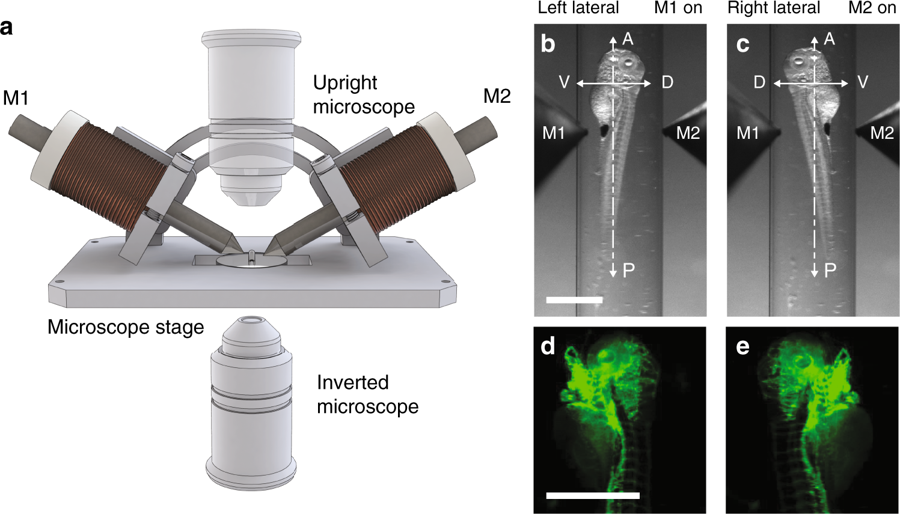
Fig. 2 Insert for multi-view imaging on a single-view microscope. a Schematic showing the insert holding two electromagnets (M1 and M2) on a microscope stage. b–e Bright-field (b, c) and fluorescence (d, e) images of a 5dpf Tg(kdrl:GFP) zebrafish larva rotated about its anterior-posterior axis by providing power to electromagnet M1 and M2, respectively. Scale bar, 1 mm
Acknowledgments
This image is the copyrighted work of the attributed author or publisher, and
ZFIN has permission only to display this image to its users.
Additional permissions should be obtained from the applicable author or publisher of the image.
Full text @ Nat. Commun.