
|
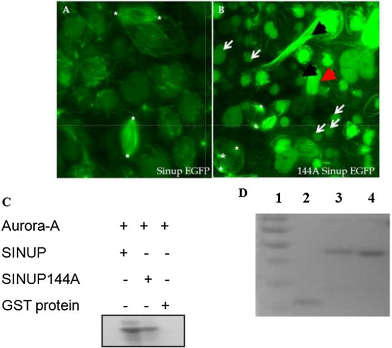
Fig. 3 Effect of misforced expression of sinup on spindle organization. (A) sinup-EGFP-injected embryo. (B) sinup144A-EGFP-injected embryo. The embryos were scanned at the sphere stage with a confocal microscope. The embryos injected with sinup144A-EGFP showed disruption in the mitotic spindle organization, orientation, and polarity of the spindle pole. Multipolar spindles were labeled with white star. (C) In vitro phosphorylation assay of Sinup144A recombinant protein. Phosphorylation of Sinup144A by Aurora A was greatly reduced in comparison with that of wild-type Sinup. (D) Purified recombinant proteins were stained with Coomassie brilliant blue on SDS-PAGE gel. 1: Marker (Fermentas #SM0671), 2: GST protein, 3: Sinup-GST, 4: Sinup144A-GST.