
Image
|
Figure Caption
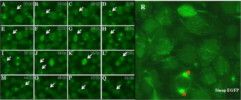
Fig. 1 Sinup is localized in the centrosomes and mitotic spindles: sinup-EGFP-injected embryos were scanned with a confocal microscope with animal pole view. (A–Q) Sinup was localized in the mitotic spindle organization throughout the cell division. (R) Magnified expression domain of sinup transcripts. Asterisks indicate centrosome and arrow indicates microtubules.
Acknowledgments
This image is the copyrighted work of the attributed author or publisher, and
ZFIN has permission only to display this image to its users.
Additional permissions should be obtained from the applicable author or publisher of the image.
Full text @ Animal Cells Syst (Seoul)