
|
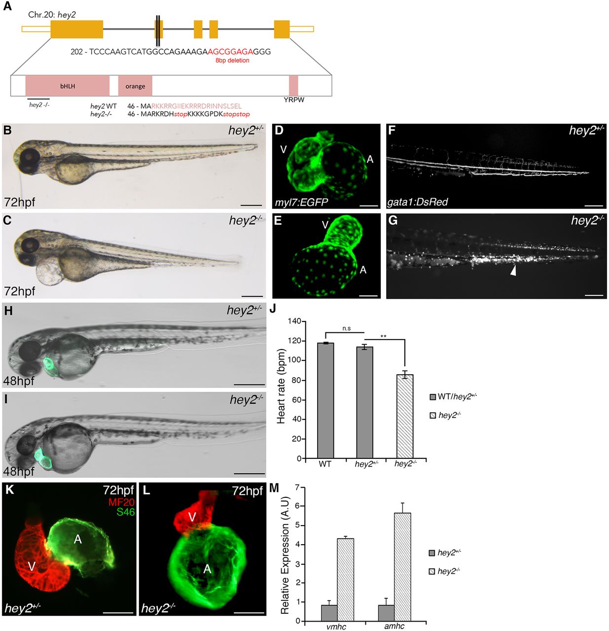
Fig. 1
Cardiovascular defects are observed in the absence of Hey2 function. (A) Schematic representation of the hey2hsc25-null allele generated through CRISPR/Cas9-mediated genome editing. Red lettering shows 8 bp deleted sequence. Protein sequence shows production of premature stop codon at the beginning of exon 2. (B,C) Bright-field images of a sibling control and a hey2hsc25 mutant embryo at 72 hpf. (D,E) Confocal images of Tg(myl7:EGFP) hearts in control (D) and hey2 mutant (E) embryos at 72 hpf. (F,G) Fluorescent images of Tg(gata1:DsRed) showing normal blood flow in controls at 72 hpf (F) and lack of blood flow leading to the accumulation of blood cells in hey2 mutants (G, arrowhead). (H,I) Bright-field images of Tg(myl7:EGFP) in hey2 heterozygous (H) and mutant (I) embryos at 48 hpf. (J) Heart rate analysis represented as beats per minute (bpm) at 48 hpf (N=3, n=4). (K,L) MF20/S46 immunofluorescence imaging at 72 hpf in hey2 heterozygous (K) and mutant (L) embryos. A, atrium; V, ventricle. (M) Quantitative RT-PCR analysis comparing amhc and vmhc gene expression in hey2 heterozygous and mutant embryos at 48 hpf (gene expression normalized to β-actin, fold difference relative to control; N=3, n=3). Data are mean±s.e.m.; **P<0.01; n.s, not significant. Scale bars: 50 µm.