
|
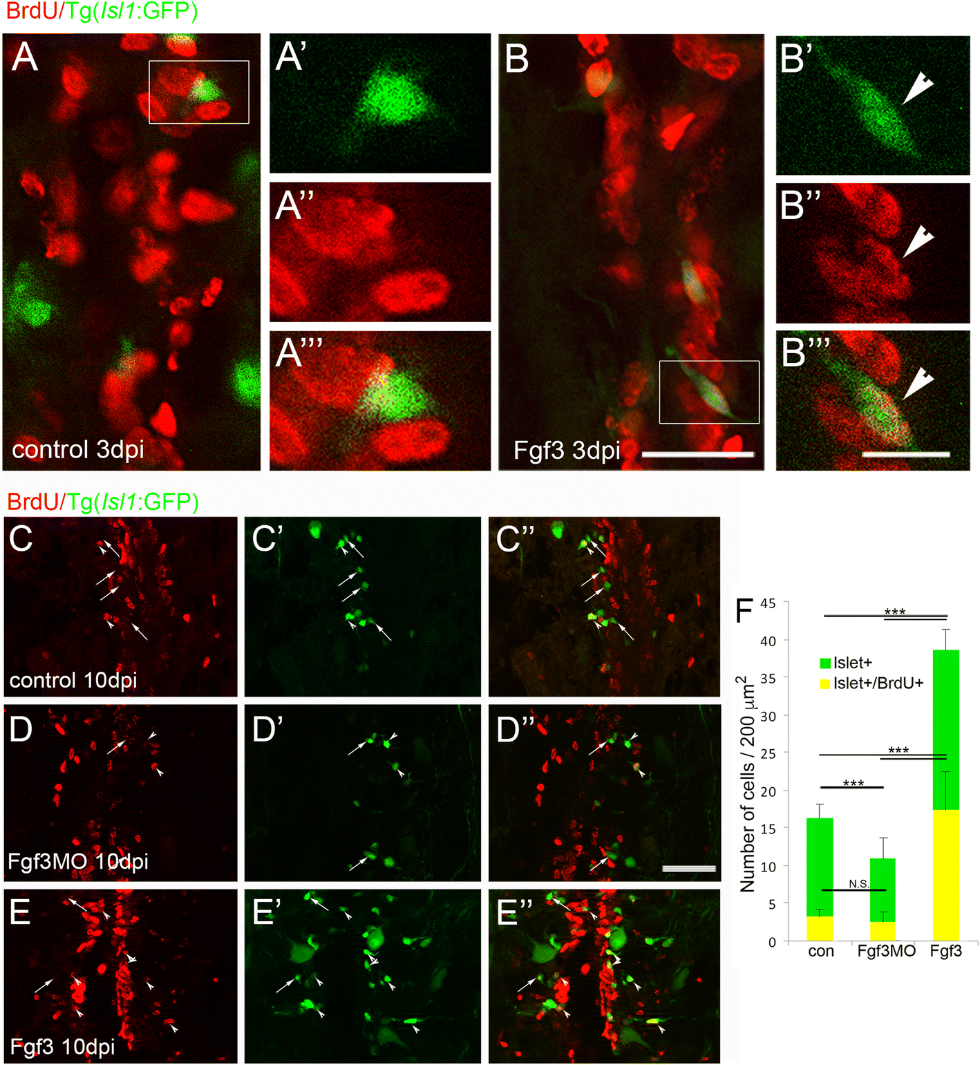
Fig. 2
Fgf3 facilitates proliferation and neurogenesis of Islet1 motor neurons after spinal cord injury. a, b Three days post spinal cord injury (dpi), very few newly generated BrdU+ (red) cells express Islet1+ (green) motor neuron marker (a), unless treated with Fgf3 for three days (B, arrowhead). White box indicates region shown at higher magnification with individual and merged channels (A’ - A”’ and B’ – B”’). (C – F) Analysis of controls at 10 dpi shows that usually only a small proportion of newly generated BrdU+ cells usually become Islet1+ motor neurons (c, f). However, treatment with Fgf3 for three days facilitates both overall proliferation (increased number of BrdU+ cells) and specifically the proportion of newly generated cells that are becoming Islet1+ motor neurons (e, f), while overall Islet+ numbers, but not the newly generated BrdU+ cohort is significantly reduced when Fgf3 signalling is inhibited (d, f). Results in C show mean ± SEM, (n = 5 fish /group) *** p < 0.001; N.S.: not significant. Scale bar in B (for A and B) is 50 μm, scale bar in B”’ (for A’ – A”’ and B’ – B”’) is 10 μm and scale bar in F” (for D - F is 100 μm)