
|
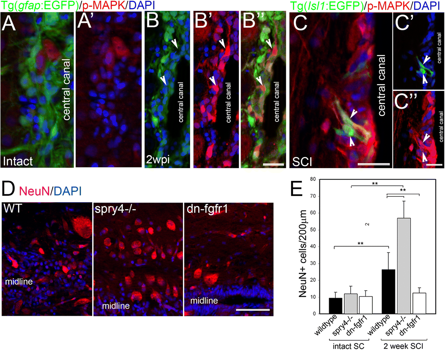
Fig. 1
Fgf signalling increases neurogenesis after spinal cord injury. a Micrographs through intact non-injured adult zebrafish spinal cord show weak p-MAPK expression. b Micrographs through adult zebrafish spinal cord two weeks post injury (wpi) shows p-MAPK levels upregulated particularly in non-radial glia GFAP negative neurons at the central canal at the lesion site (arrows, B) (n = 5) some of which are Islet1 positive (c). d, e While Fgf signalling gain (spry4−/−) or loss (Tg(hsp70l:dn-fgfr1-EGFP) has no effect on NeuN+ neurons in intact spinal cord (SC), two weeks after injury, the significant increase in NeuN+ neurons in WT can be further increase with Fgf signaling gain and abolished with Fgf signaling loss. Graphs shows mean ± SEM, (n = 6 fish /group) ** p < 0.01. Scale bars in A, B and C are 25 μm, scale bar in D is 50 μm