
|
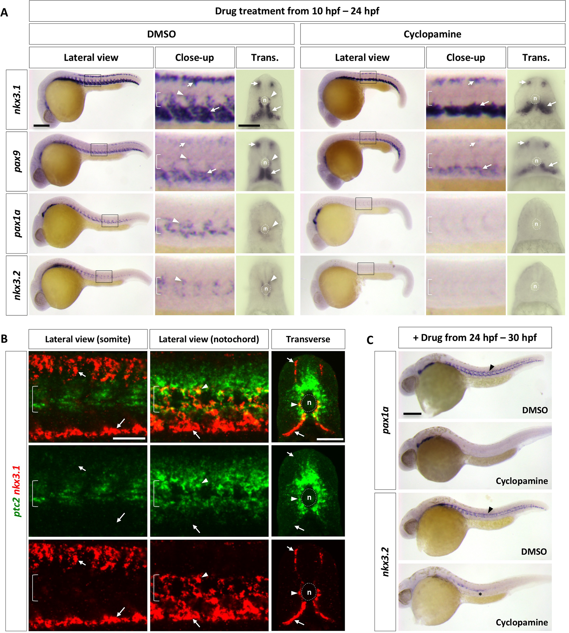
Fig. 2
Regulation of the zebrafish sclerotome by Hh signaling.
(A) Wild-type embryos were treated with either DMSO or cyclopamine between 10 hpf and 24 hpf, and stained for the expression of nkx3.1, pax9, pax1a, and nkx3.2. The expression of nkx3.1 and pax9 in dorsal and ventral sclerotome domains (short and long arrows, respectively) remained the same upon cyclopamine treatment, while expression of all sclerotome markers was absent in sclerotome derived notochord associated cells (arrowheads). The notochord (n) is indicated by brackets in lateral views and dotted lines in transverse views. n = 45 embryos per condition. (B) Wild-type embryos at 24 hpf were co-labeled with ptc2 (green) and nkx3.1 (red). nkx3.1 expressing dorsal and ventral sclerotome domains (short and long arrows, respectively) do not express ptc2, whereas nkx3.1 expressing cells surrounding the notochord (arrowheads) are positive for ptc2. The confocal plane of the somite shows dorsal and ventral sclerotome domains, while the optical slice near the notochord reveals notochord associated cells and the ventral sclerotome. n = 15 embryos. (C) Wild-type embryos were treated with either DMSO or cyclopamine between 24 hpf and 30 hpf, and stained for the expression of pax1a and nkx3.2 at 30 hpf. pax1a and nkx3.2 expression was absent in sclerotome derived notochord associated cells (arrowheads) upon treatment of cyclopamine. Note that nkx3.2 expression in migrating lateral line cells (asterisk) was not affected. n = 30 embryos per staining. Scale bars: (A, C) 200 μm in lateral views, 50 μm in transverse views; (B) 50 μm.