
|
Fig. S5
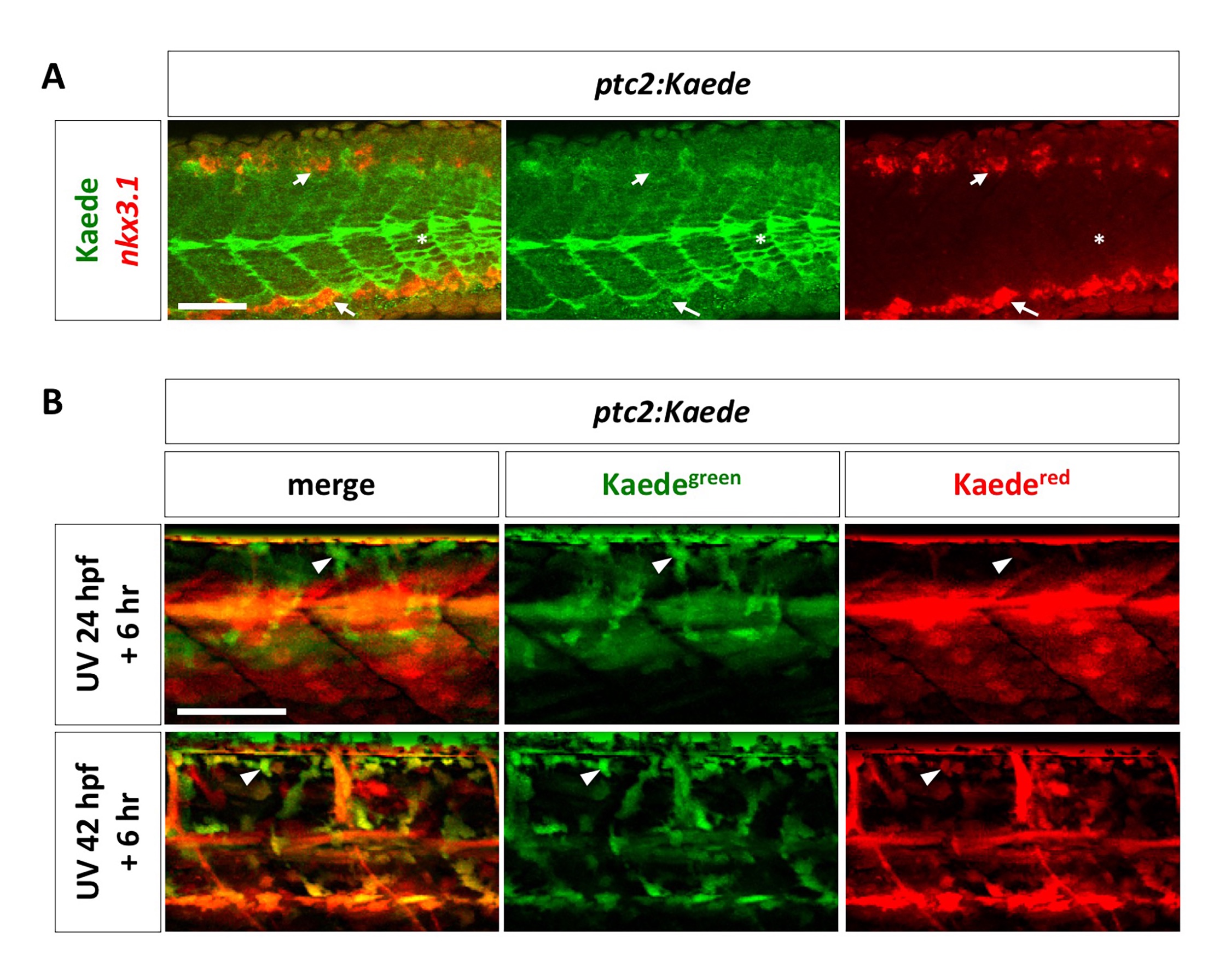
Analysis of Hh response in the sclerotome.
(A) ptc2:Kaede transgenic embryos were co-labeled using the nkx3.1 probe (red) and the Kaede antibody (green) at 24 hpf. Neither the dorsal sclerotome domain (short arrows) nor the ventral sclerotome domain (long arrows) labeled by nkx3.1 have overlapping expression with Kaede. ptc2:Kaede expression in slow muscle fibers are indicated by asterisks. n = 15 embryos. (B) ptc2:Kaede embryos were photoconverted at 24 hpf or 42 hpf, and imaged 6 hours later (top and bottom panel, respectively). Kaedegreen signal represents “new” signaling activity within the 6-hour time window, whereas Kaedered signal represents “old” signaling that occurs before the time of photoconversion. ptc2:Kaede expression is present in presumptive sclerotome derived notochord associated cells (arrowheads) at both 30 hpf and 48 hpf. n = 4 embryos per time point. Scale bars: 50 μm.