
|
Fig. 4
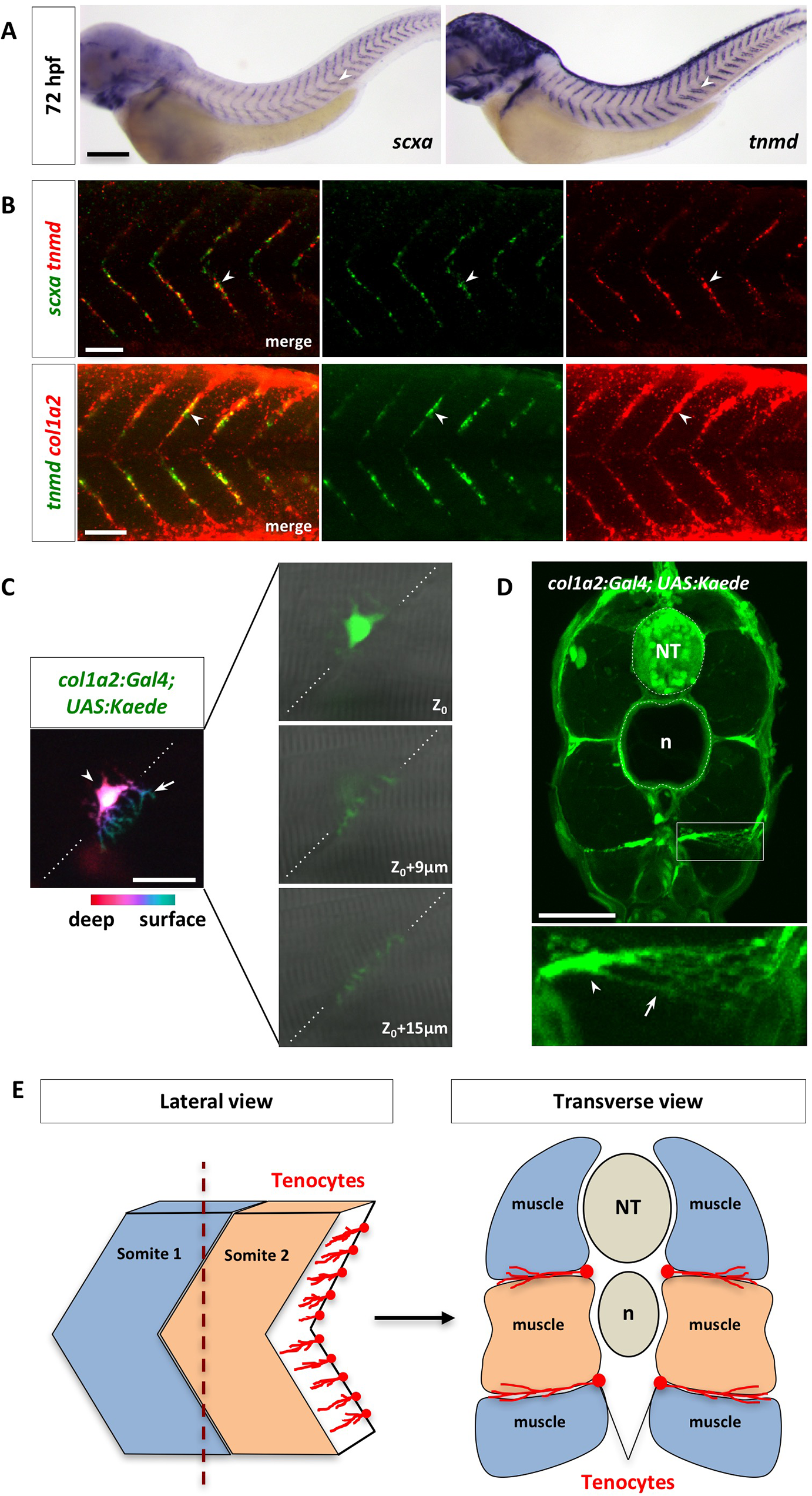
Characterization of zebrafish axial tenocytes.
(A) Expression of tenocyte markers scxa and tnmd in wild-type embryos at 72 hpf. Both scxa and tnmd are expressed along the V-shaped myotendinous junction (MTJ) between somites (arrowheads). The complete time course of expression is shown in S6 Fig. n = 30 embryos per staining. (B) Wild-type embryos at 72 hpf were co-labeled with scxa and tnmd (green and red in the top panel, respectively), or tnmd and col1a2 (green and red in the bottom panel, respectively). Overlapping expression between scxa, tnmd, and col1a2 were observed in tenocytes along the MTJ (arrowheads). n = 60 embryos per staining. (C) Mosaic col1a2Kaede embryos at 3 dpf were selected to image individual Kaede+ tenocytes. Color coded depth projection of a single col1a2Kaede tenocyte is shown on the left. The tenocyte cell body (arrowhead) is located deeper (magenta) along the MTJ (dotted lines), whereas its cellular processes (arrow) are projected towards the surface (cyan). Three individual confocal slices of the same tenocyte are shown on the right. The cell body is found near the medial edge of the somite (Z0) and its cellular projections extend through the MTJ (Z0 + 9 μm and Z0 + 15 μm) towards the lateral surface. n = 25 embryos. (D) Cross section view of col1a2Kaede embryos at 5 dpf. An expanded view of a tenocyte in a boxed region is shown at the bottom. The cell bodies of tenocytes (arrowhead) are located at the medial edge of the MTJ, and they extend long cellular projections (arrow) towards the surface of the embryo. n = 28 tenocytes from 15 embryos. (E) Schematic representations of lateral and transverse views of tenocyte organization along the somite boundaries. NT: neural tube; n: notochord. Scale bars: (A) 200 μm; (B) 50 μm; (C) 20 μm; (D) 50 μm.