
|
Fig. S5
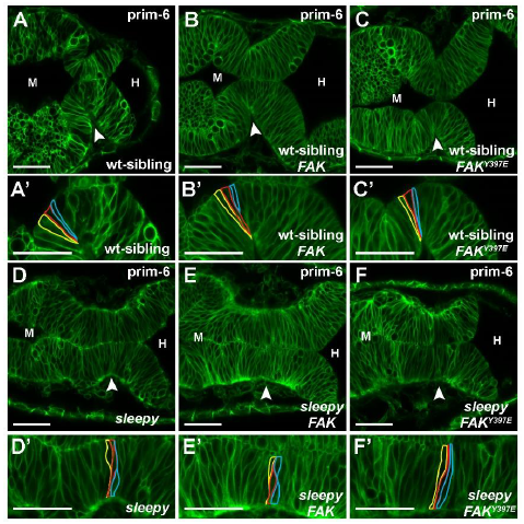
Expression of human FAK does not rescue the laminin mutant basal constriction phenotype. (A-F’) Live confocal images showing the MHB region of prim-6 embryos injected with mGFP (A,D), mGFP + wt FAK mRNA (B,E), mGFP + FAK-Y397E (C,F). (A-C’) sleepy (slym86) (Schier et al., 1996) heterozygous sibling or wild-type sibling embryos showing normal basal constriction at the MHBC. (D-F’) sleepy mutants showing defects in basal constriction both without and with the co-injection of human wild-type FAK and human phosphomimetic FAKY397E mRNA. Representative images from 3 independent experiments with n>3 for each condition. (A’-F’) Magnifications of the neuroepithelium shown in A-F with individual cells outlined at the MHBC. Arrowheads indicate MHBC. M, midbrain. H, hindbrain. Scale bars: 35 m. Biology Open (2018): doi:10.1242/bio.034520: Supplementary information Biology Open • Supplementary information