
|
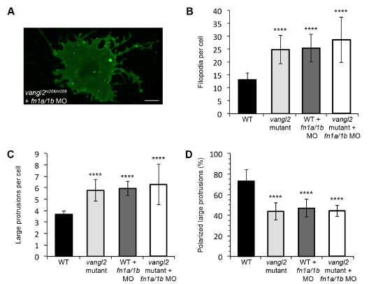
Fig. S10
Knockdown of fibronectin in vangl2 mutant embryos. (A) Time-lapse confocal image of late gastrula stage cell expressing memGFP. vangl2 mutant embryo injected with fn1a/1b morpholinos (MO). (B,C) Quantitation of the total numbers of membrane protrusions formed by vangl2 mutant embryos injected with fn1a/1b MO (n=5 cells, 3 embryos). The data from wild type (WT), vangl2 mutant embryos, and wild-type embryos injected with fn1a/1b MO are shown for comparison. (D) Quantitation of the total percentage of polarized large protrusions. Average values are shown ± standard deviation. ****P<0.0001; P values are versus wild type; one-way ANOVA significance test followed by Tukey HSD post-hoc tests. Scale bar, 5 μm.