
Image
|
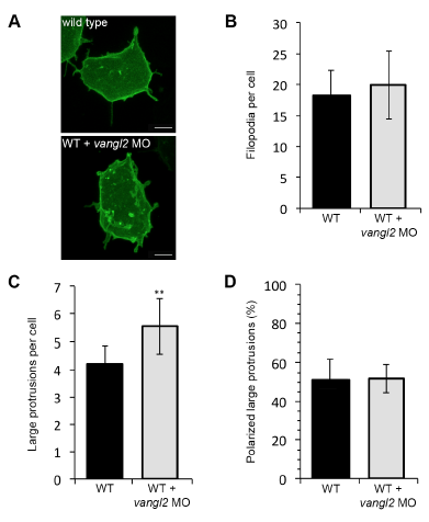
Figure Caption
Fig. S7
Wild type and vangl2 morphant membrane protrusive activity at midgastrulation. (A) Time-lapse confocal images of 80% epiboly epiblast cells expressing memGFP. WT, wild type. (B,C) Quantitation of the total numbers of membrane protrusions formed by wild type (n=9 cells, 7 embryos) and wild-type embryos injected with vangl2 morpholino (MO) (n=10 cells, 6 embryos). (D) Quantitation of the total percentage of polarized large protrusions. Average values are shown ± standard deviation. **P<0.01; P values are versus wild type; two-tailed unpaired t-test. Scale bars, 5 μm.
Acknowledgments
This image is the copyrighted work of the attributed author or publisher, and
ZFIN has permission only to display this image to its users.
Additional permissions should be obtained from the applicable author or publisher of the image.
Full text @ Development