
Image
|
Figure Caption
Fig. S5
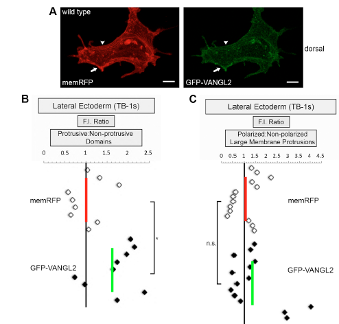
GFP-VANGL2 localization in protrusive and non-protrusive membrane domains. (A) Lateral ectodermal cell labeled with memRFP and GFP-VANGL2. Arrows denote a forming non-polarized large protrusion. Arrowheads show a non-protrusive membrane domain. memRFP and GFP-VANGL2 fluorescence intensity (F.I.) ratios for (B) protrusive/non-protrusive membrane domains (n=9 protrusions, 6 embryos) and (C) polarized/non-polarized large protrusions (n=14 protrusions, 5 embryos). Vertical red and green lines indicate the averages. *P<0.05, n.s. not significant; two-tailed paired t-test. Scale bars, 5 μm.
Acknowledgments
This image is the copyrighted work of the attributed author or publisher, and
ZFIN has permission only to display this image to its users.
Additional permissions should be obtained from the applicable author or publisher of the image.
Full text @ Development