
|
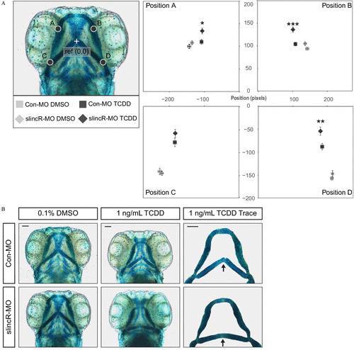
Fig. 4
Effect of 1ng/mL TCDD on the developing jaw after treatment of control and slincR morphants. Control (Con-MO) and slincR morphants (slincR-MO) were developmentally exposed to 0.1% DMSO or 1ng/mL TCDD, and the cartilage was stained and measured at 72hpf . (A) A morphometric system was used to measure the position and length of landmark structures in the developing jaw. The position of jaw structures representing junctions between Meckel’s and palatoquadrate cartilages (point A and B) and representing junctions between the hyosymplectic and ceratohyal cartilages (point C and D) was measured relative to a reference point as shown. Statistical significance was determined by a modified two-way ANOVA with a Tukey post hoc test. Morphometric values represent mean±standarderrorofthemean . ( n=9–10 individual 72-hpf zebrafish; p<0.05=* , p<0.01=** , p<0.001=*** ). The asterisk (*) indicates statistical significance between slincR-MO and Con-MO samples exposed to TCDD. (B) Representative images of the cartilage structures in control and slincR morphants treated with DMSO or TCDD. The arrow in the TCDD Trace panel points to the junction between the hyosymplectic and ceratohyal cartilages (point C and D). The bar in the top right corner indicates 100μM .