
|
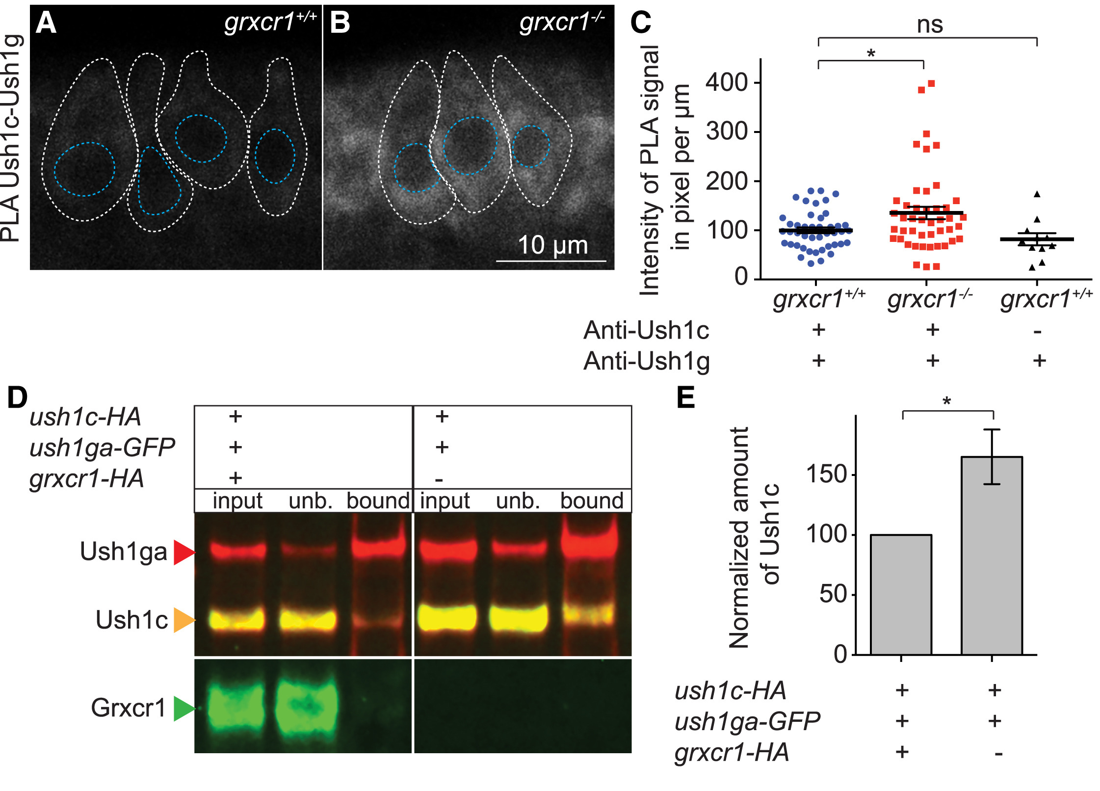
Fig. 7
Grxcr1 Destabilizes Ush1c-Ush1ga Interactions
(A and B) Proximity ligation assay for Ush1c and Ush1g in the anterior maculae of grxcr1+/+ (A) and grxcr1−/− larvae (B) at 5 dpf.
(C) Quantification of Ush1c-Ush1g interactions in grxcr1+/+ (n = 44 larvae), grxcr1−/− larvae (n = 45 larvae), and grxcr1+/+ larvae that were incubated with only one primary antibody (control, n = 11 larvae). Data were normalized to grxcr1+/+. Individual hair cells are outlined in white. Nuclei are outlined in blue.
(D) MDCK cells were transfected with ush1c-HA, ush1ga-GFP, and grxcr1-HA (+) or not (−). Extracted proteins were immunoprecipitated with an anti-GFP antibody and blotted using anti-Ush1c, anti-GFP, and anti-HA antibodies.
(E) Quantification of the Ush1c level in bound fractions in the presence or absence of Grxcr1 (n = 4 experiments). Data were normalized to input and are presented as a ratio amount of normalized Ush1c in the presence of Grxcr1 over the amount of normalized Ush1c in the absence of Grxcr1.
Data are represented as mean ± SEM. ∗p < 0.05.