
Image
|
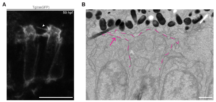
Figure Caption
Fig. S2
Formation of processes in the growing IS of maturing PRCs. A: Visualisation, by confocal imaging, of retinal sections of embryos at 59 hpf expressing the Tg(rasGFP) construct (plasma membrane marker). Processes (arrowhead) appear at the same time as the IS becomes visible and are still visible when OSs form. B: Electron micrograph of a section through the PRC layer in zebrafish embryo aged 55 hpf. The magenta dashed line highlights a process extending from the growing IS (magenta arrow). Scale bars: A: 10 µm, B:1 µm.
Acknowledgments
This image is the copyrighted work of the attributed author or publisher, and
ZFIN has permission only to display this image to its users.
Additional permissions should be obtained from the applicable author or publisher of the image.
Full text @ Biol. Open