
Image
|
Figure Caption
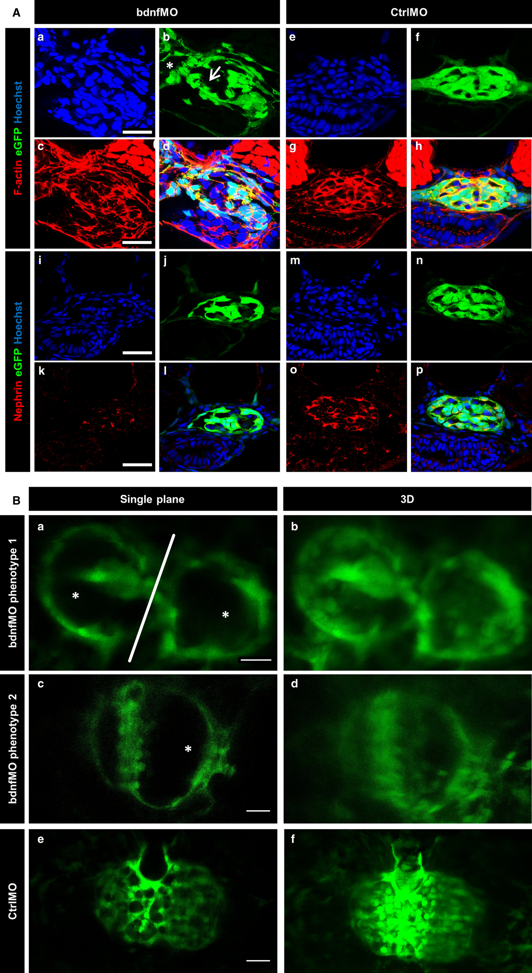
Fig. 6
Bdnf knockdown in zebrafish larvae leads to morphological changes in the glomerulus and the down‐regulation of podocyte marker proteins. BdnfMO and CtrlMO were injected in ET eggs expressing eGFP specifically in podocytes. Cryosections were counterstained for F‐actin by phalloidine (red) and nuclei by Hoechst (blue) (A; a‐h). bdnfMO‐treated larvae show an enlarged glomerular tuft (A; b arrow) and Bowman's space (A; b asterisk) compared with the normal morphology of the CtrlMO‐treated larvae (A; e‐h). Counterstaining of the slit diaphragm protein nephrin (red) and nuclei by Hoechst (blue, A; i‐p) reveals a down‐regulation of nephrin due to bdnfMO treatment (A; k) compared to CtrlMO‐treated larvae (A; o). [Scale bars = 20 μm] In vivo microscopy reveals 2 different phenotypes of bdnfMO‐treated larvae. Phenotype 1 is characterized by unfused glomeruli (B; a white line and b), a reduced number of podocytes and a dilatation of Bowman's space and the glomerular tuft (B; a asterisks). The second phenotype is characterized by the absence of podocyte major processes, a reduced podocyte number and a dilatation of Bowman's space and the glomerular tuft (B; c asterisk and d). CtrlMO‐injected larvae show a normal glomerular morphology with well‐shaped major processes (B; e and f). [Scale bars = 20 μm]
Figure Data
Acknowledgments
This image is the copyrighted work of the attributed author or publisher, and
ZFIN has permission only to display this image to its users.
Additional permissions should be obtained from the applicable author or publisher of the image.
Full text @ J. Cell. Mol. Med.