
|
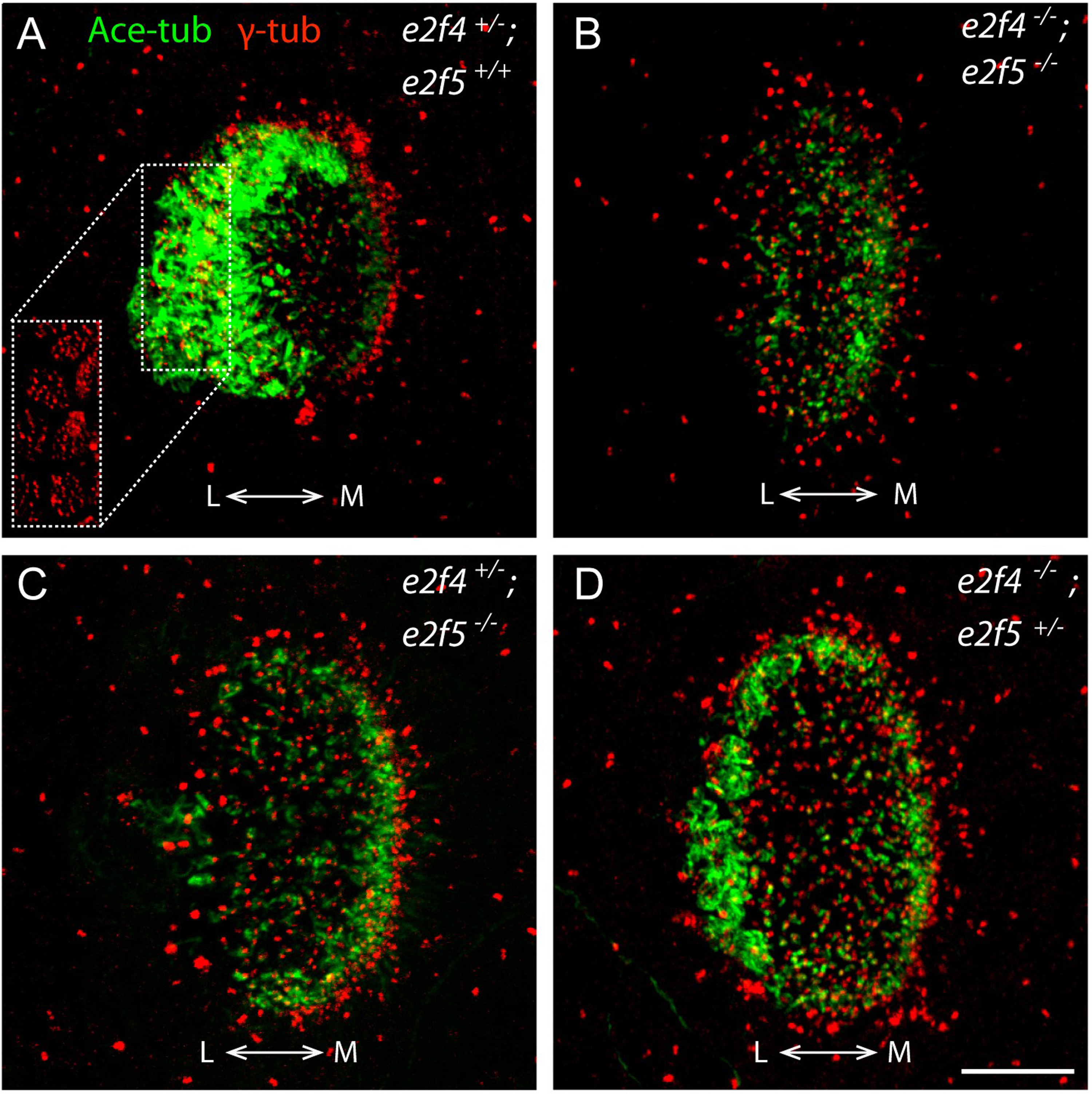
Fig. 3
Both e2f4 and e2f5 contribute to MCC formation at the nasal placode. A: Thick layers of motile cilia bundles (labelled by anti-acetylated α-tubulin (Ace-tub)) were featured prominently on the lateral side of e2f4+/- nasal placodes. MCCs can be identified by patches of densely packed basal bodies (labelled by anti-gamma-tubulin (γ-tub)), a subset of which is featured in the single channel inset (white dotted rectangle, image scale unchanged). B: e2f4-/-; e2f5-/- double homozygous mutants show a complete lack of MCCs. C: MCCs are essentially absent in the nasal placodes of e2f4+/-; e2f5-/- embryos. D: The nasal placode MCC layers are present but much thinner in e2f4+/-; e2f5-/- mutants than that in wild-type or heterozygous siblings. All embryos were siblings from an inter-crosses of e2f4+/-; e2f5+/- adult mutants and were fixed for immunostaining at 72 hpf. L: lateral; M: medial. Scale bar = 10 µm.
Reprinted from Developmental Biology, 443(2), Chong, Y.L., Zhang, Y., Zhou, F., Roy, S., Distinct requirements of E2f4 versus E2f5 activity for multiciliated cell development in the zebrafish embryo, 165-172, Copyright (2018) with permission from Elsevier. Full text @ Dev. Biol.