
|
Fig. 2
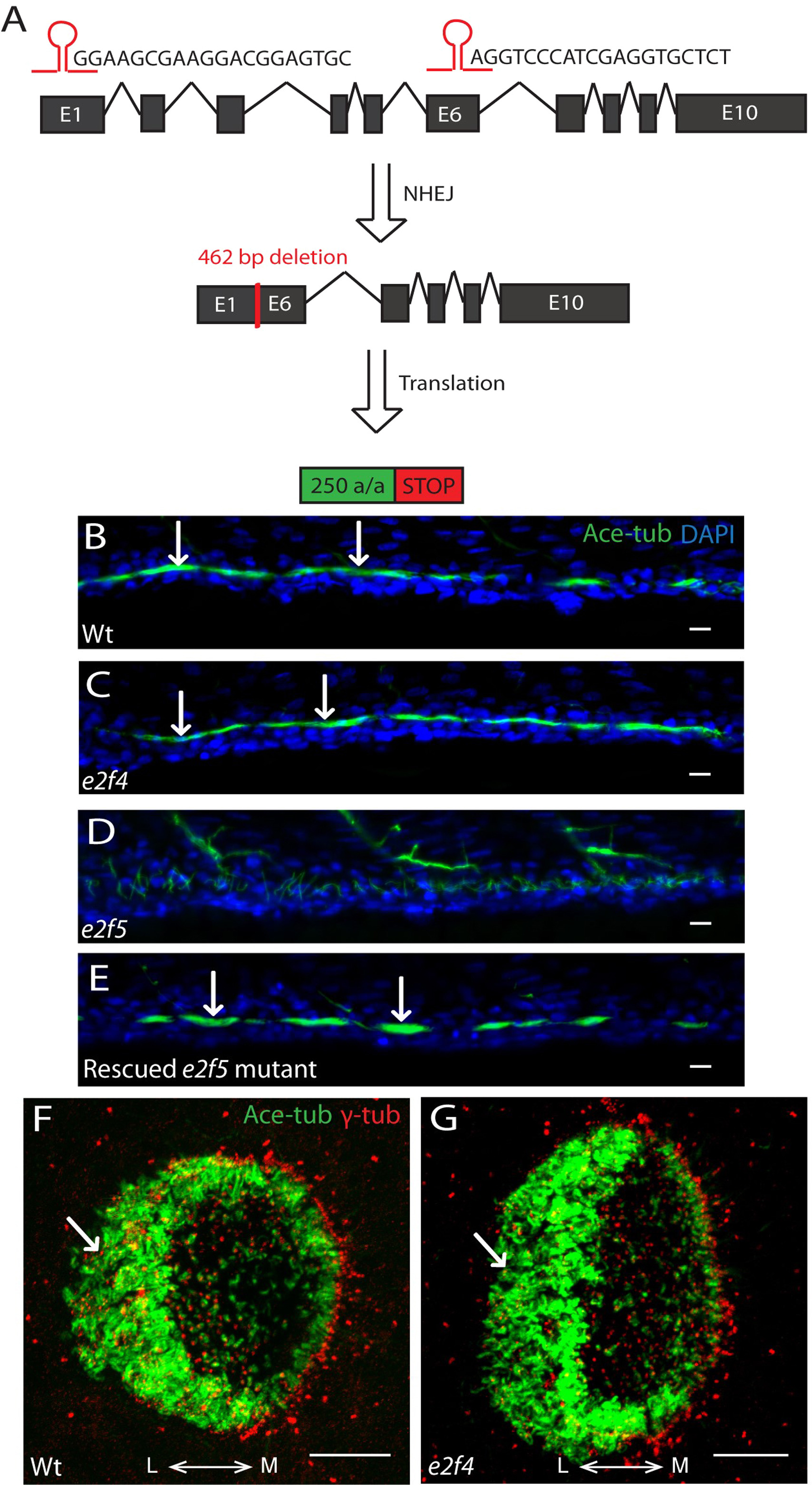
E2f4 is dispensable for MCC formation. A: Schematic showing CRISPR/Cas9-mediated genome editing at the zebrafish e2f4 locus using two guide RNAs targeting exon 1 and exon 6. After NHEJ, F1 carriers contained a 462-bp deletion between exon 1 and exon 6. This resulted in a 154-amino acid in-frame deletion. B: Kidney tubule of a 48 hpf wild-type embryo populated with clusters of MCCs (arrows). C: MCCs are unaffected in the kidney tubule of an e2f4 homozygous mutant (arrows). Acetylated-tubulin (Ace-tub; green), DAPI (blue). Scale bars: 10 µm. D: Kidney tubule of an e2f5 mutant embryo completely devoid of MCCs. E: Rescued MCCs in the kidney tubule of an e2f5 mutant by over-expression of E2f4 (arrows). Acetylated-tubulin (Ace-tub; green), DAPI (blue). Scale bars= 10 µm. F: MCCs around the lateral rim of a nasal placode (arrow) of a wild-type embryo at 72 hpf. G: Nasal placode MCCs are unaffected in an e2f4 mutant (arrow). Acetylated-tubulin (Ace-tub; green), γ-tubulin (γ-tub; red). L: lateral; M: medial. Scale bars = 10 µm. (For interpretation of the references to color in this figure legend, the reader is referred to the web version of this article).
Reprinted from Developmental Biology, 443(2), Chong, Y.L., Zhang, Y., Zhou, F., Roy, S., Distinct requirements of E2f4 versus E2f5 activity for multiciliated cell development in the zebrafish embryo, 165-172, Copyright (2018) with permission from Elsevier. Full text @ Dev. Biol.