
|
Fig. S10
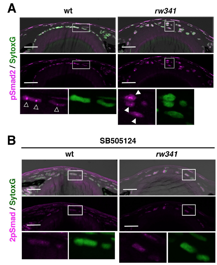
SB505124 treatment inhibits TGF-β signaling in zebrafish lens epithelium
(A) Labeling of 5 dpf wild-type and rw341 mutant lenses with anti-pSmad2 antibody (magenta) and Sytox Green (green). Upper panels show lens epithelium. Middle panels indicate the magenta channel. Bottom panels show higher magnifications of squares indicated in the upper/middle panels. In wild-type lenses, small dotted pSmad2 signals are observed in lens epithelial cell nuclei (open arrowheads). In rw341 mutants, pSmad2 signals increased throughout the whole region of lens epithelial cell nuclei (filled arrowheads).
(B) Labeling of 5 dpf SB505124-treated wild-type and rw341 mutant lenses with anti-pSmad2 antibody (magenta) and Sytox Green (green). Upper panels show lens epithelium. Middle panels indicate the magenta channel. Bottom panels show higher magnification of squares indicated in the upper/middle panels. SB505124 treatment markedly reduces the intensity of pSmad2 signals in both wild type and rw341 mutant lens epithelium, confirming that this concentration of SB505124 effectively inhibits TGF-β signaling in lens epithelium. Scale bars: 20 μm (A, B).