
Fig. 2
- ID
- ZDB-IMAGE-181121-22
- Publication
- Chen et al., 2018 - Fertility Enhancement but Premature Ovarian Failure in esr1-Deficient Female Zebrafish.
- All Figures
- Figures for Chen et al., 2018
|
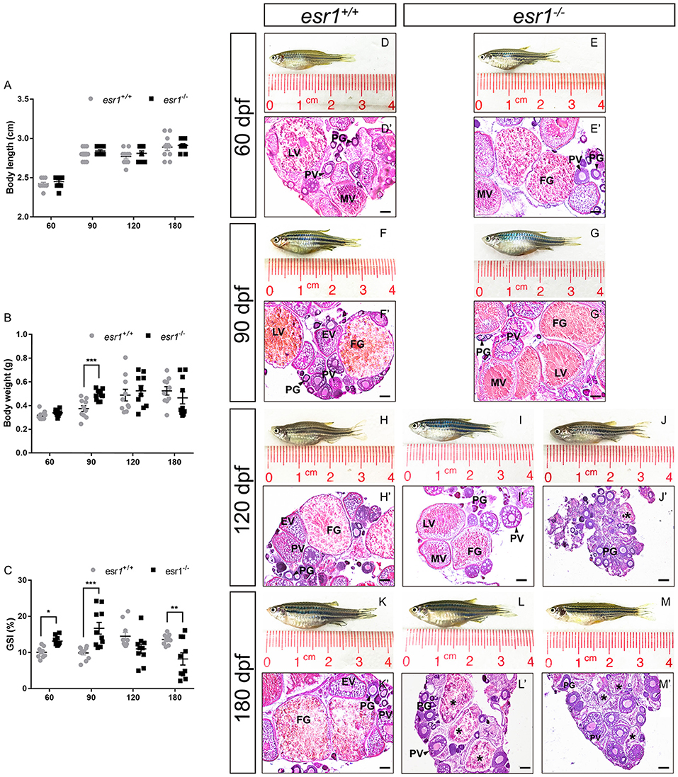
Fig. 2
Morphological and histological analyses of esr1 mutant female zebrafish. (A) Body length of esr1+/+ and esr1−/− female zebrafish. (B) Body weight of esr1+/+ and esr1−/− female zebrafish. (C) GSI of esr1+/+ and esr1−/− female zebrafish. (D–M) Gross morphology of female zebrafish. (D′-G′) Ovaries of esr1+/+ and esr1 mutant were normal at 60 and 90 dpf. (H′-M′) Part of esr1 mutant ovaries of at 120 dpf and all esr1 mutant ovaries were degenerated by the 180 dpf. In these degenerated ovaries, MV and FG follicles were absent and PG follicles, PV follicles and atretic follicles (asterisk) were found. PG, primary growth follicle; PV, pre-vitelligenic follicle; EV, early-vitelligenic follicle; MV, middle-vetelligenic follicle; LV, late-vitelligenic follicle; FG, full grown follicle; *, atretic follicle.