
|
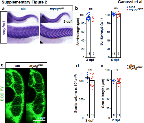
Fig. S2
Myogenin mutant has normal smyhc1 expression and myogfh265 has no growth defect.
a. ISH shows that at 2 dpf smyhc1 mRNA level is indistinguishable in myogkg125 mutant and sibs, but mutant embryos have a reduced extent of somitic muscle (red brackets). b. Quantification of somite length at 3 and 5 dpf. c. Optical cross sections of myogfh265 mutants and sibs stained with BODIPY. d. Quantitation of somite volume measured in “c”. e. Somite length measurement shows that mutants and sibs are comparable. t-test, ns= not significant. Replicate numbers are given in Supplementary Table 2. Bars = 50 μm.