
|
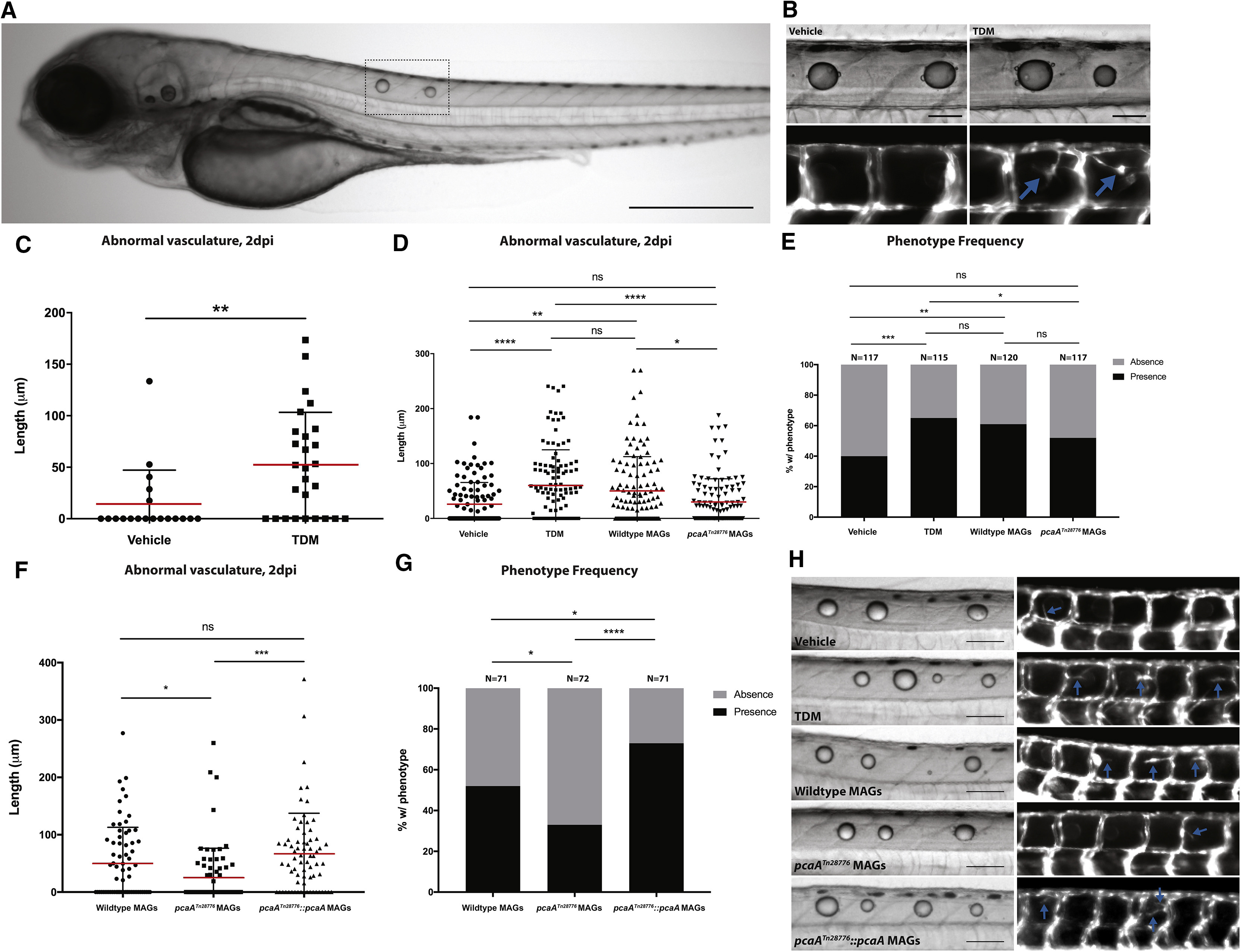
Fig. 2
Purified TDM and Mycolic Acid Glycolipids from Wild-Type Bacteria but Not pcaA Mutants Elicit Robust Angiogenesis
(A) Representative image of 2-day post-injection larvae injected dorsally with TDM. Dotted line indicates injection site and droplet coalescence. Scale bar, 500 μm.
(B) Representative images of injections of either vehicle (left) or TDM (right) into Tg(kdrl:EGFP) animals. Blue arrows indicate abnormal vasculature (lower panels). Scale bars, 100 μm.
(C) Quantification of abnormal vasculature induced by TDM or vehicle alone. Representative of three experiments. ∗∗p < 0.01, Student's t test.
(D) Quantification of abnormal vasculature induced by MAGs isolated from wild-type or pcaA mutant M. marinum, with vehicle alone and TDM as negative and positive controls, respectively. Data from three experiments combined. ∗p < 0.05; ∗∗p < 0.01; ∗∗∗∗p < 0.0001; one-way ANOVA with Tukey's multiple comparison post-test.
(E) Frequency of presence or absence of abnormal vasculature. ∗p < 0.05; ∗∗p < 0.01; ∗∗∗p < 0.001; Fisher's exact test.
(F) Quantification of the length of abnormal vasculature induced by MAGs (5 mg/mL) isolated from pcaATn28776:pcaA. MAGs from wild-type and pcaATn28776 included as positive and negative controls. Data from three experiments combined. ∗p < 0.05; ∗∗∗p < 0.001; one-way ANOVA with Tukey's multiple comparison post-test.
(G) Frequency of presence or absence of abnormal vasculature. ∗p < 0.05; ∗∗∗∗p < 0.0001; Fisher's exact test.
(H) Representative images of larvae injected with vehicle, TDM, wild-type MAGs, MAGs isolated from pcaATn28776, and MAGs isolated from pcaATn28776:pcaA. Blue arrows indicate abnormal vasculature. Scale bars, 100 μm.