
|
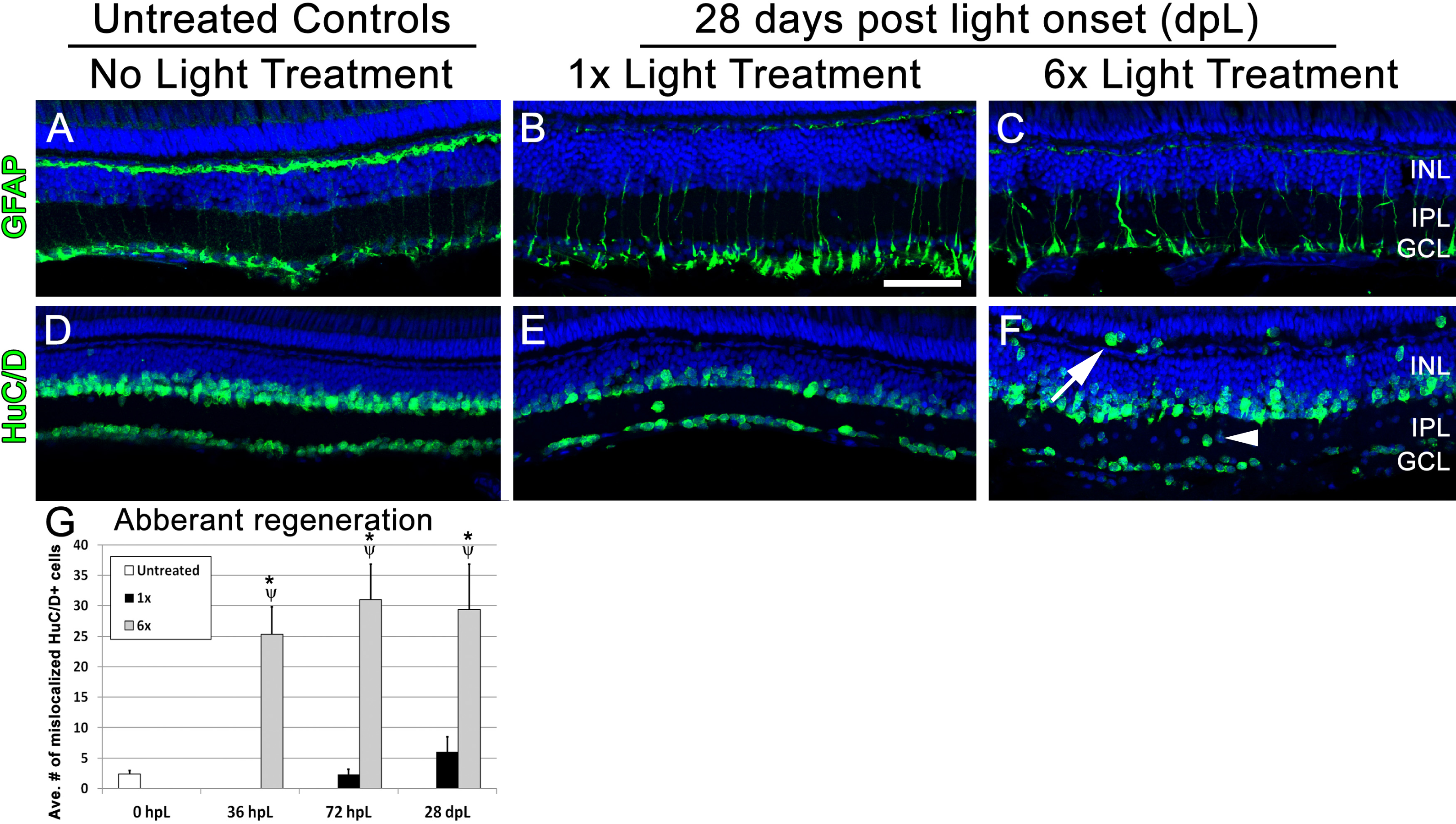
Fig. 5
Following multiple rounds of regeneration, retinas exhibit abnormal localization of inner retinal neurons.(A–F) Retinal sections collected at 28 dpL immunolabeled with anti-GFAP and anti-HuC/D to show Müller glia and inner retinal neurons, respectively, in untreated control (No Light Treatment), experimental control (1×Light Treatment) and experimental retinas (1×Light Treatment). Nuclei are stained in blue with TO-PRO-3. (A–C) Müller glia are immunolabeled with anti-GFAP. (D–F) Amacrine and ganglion cells are immunolabeled with anti-HuC/D. Mis-localization of HuC/D-positive cells were observed in the inner plexiform layer (IPL; arrowhead) and outer retina (arrow). (G) Quantification of average numbers of HuC/D-positive cells found in the IPL and outer retina (n = 5 per group). Cells were counted over a linear distance of 300 μm on the central dorsal retina. Asterisk indicates significantly different from 0 h control; Psi symbol indicates significantly different from 1×retinas (p < 0.05). Scale bar represents 25 μm.