
|
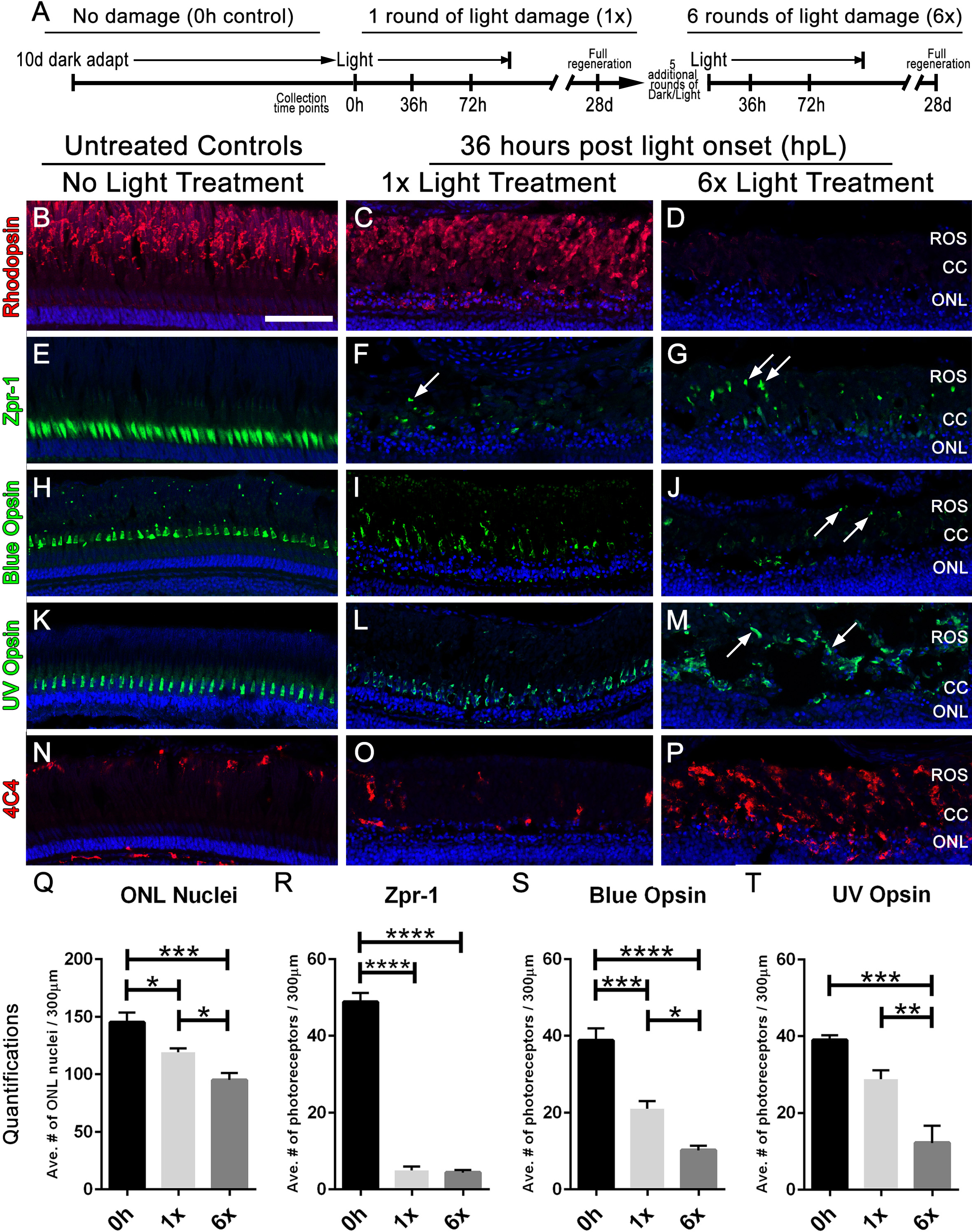
Fig. 2
Multiple rounds of light treatment leads to greater photoreceptor loss at 36 h of light treatment.(A) Experimental design used for multiple rounds of light treatment. (B–P) Retinal sections collected at 36 hpL immunolabeled with anti-rhodopsin, Zpr-1, anti-blue opsin, anti-UV opsin, and 4C4 to show the varying amounts of damage to photoreceptors in untreated control (No Light Treatment), experimental control (1×Light Treatment), and experimental retinas (1×Light Treatment). Nuclei are stained blue with TO-PRO-3. (B–D) Rod photoreceptor outer segments are immunolabeled with anti-rhodopsin (red). (E–G) Red-green double cones are immunolabeled with Zpr-1 (green). (H–J) Long single cones are immunolabeled with anti-blue opsin (green). (K–M) Short single cones are immunolabeled with anti-UV opsin (green). (N–P) Microglia/macrophages are immunolabeled with 4C4 (red). In the images taken after six rounds of light treatment, arrows point to individual cone opsins in the large debris field. (Q–T) Quantification of average numbers of photoreceptors in untreated (0h), control (1×) and experimental (1×) groups (n = 5 per group) counted over a linear distance of 300 µm on the central dorsal retina. Asterisks indicates significant differences between groups (four asterisks p < 0.0001; three asterisks p < 0.0003; two asterisks p < 0.006; one asterisk p < 0.02). Scale bar represents 25 μm.