
IMAGE
Fig. 1
- ID
- ZDB-IMAGE-181101-11
- Genes
- Antibodies
- Publication
- Kaufman et al., 2018 - rbpms2 functions in Balbiani body architecture and ovary fate
- All Figures
- Figures for Kaufman et al., 2018
Image
|
Figure Caption
Fig. 1
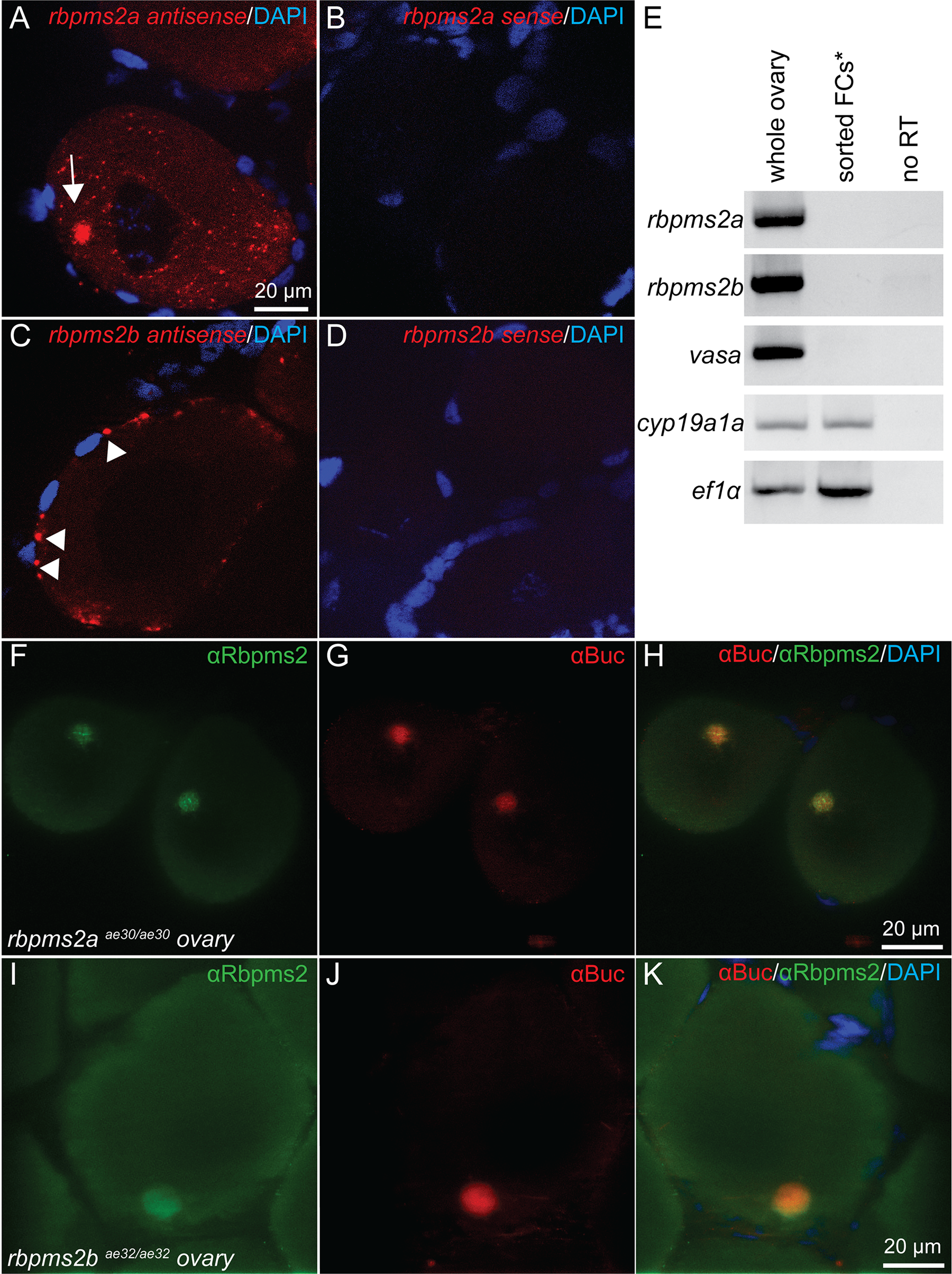
Expression of rbpms2 in oocytes.
Fluorescent in situ hybridization displaying Bb localization (arrow) of rbpms2a transcripts (A), while rbpms2b transcripts are not Bb-localized (C). Sense control probes demonstrate that rbpms2a and rbpms2b signals are specific (B,D). (E) RT-PCR on FACS-sorted granulosa and theca cells demonstrates that expression of rbpm2a and rbpms2b is germ cell specific. Antibody staining for Rbpms2 in ovaries mutant for rbpm2a (F) or rbpms2b (I) suggests both ohnologs are Bb localized where Bucky ball protein also resides (G,H,J,K).
Figure Data
Acknowledgments
This image is the copyrighted work of the attributed author or publisher, and
ZFIN has permission only to display this image to its users.
Additional permissions should be obtained from the applicable author or publisher of the image.
Full text @ PLoS Genet.