
|
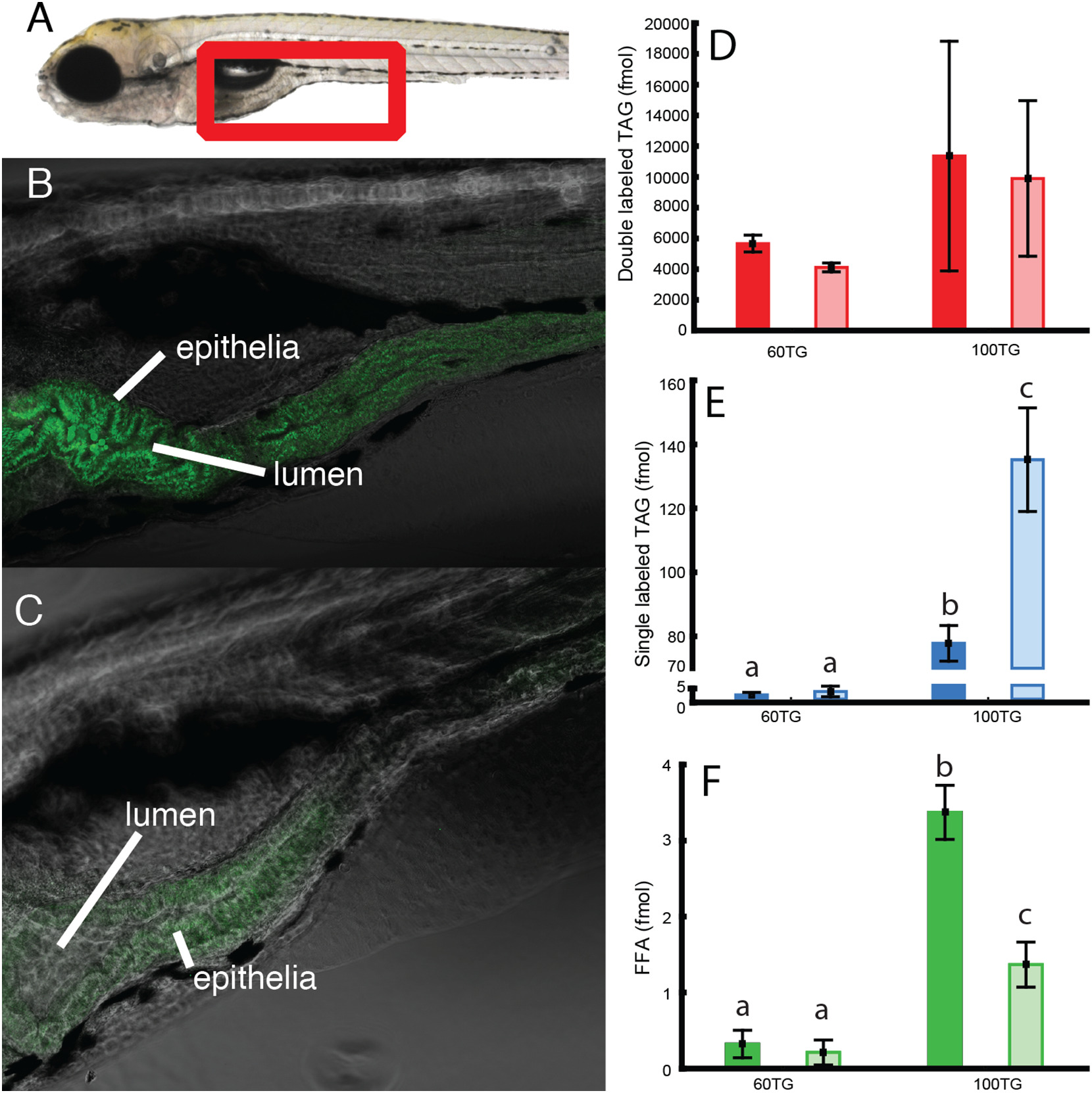
Fig. 4
Left panel: anterior intestine of zebrafish larvae (see red frame in A). Larvae in panel B were fed a 100% TG diet and larvae in panel C were fed 60% TG and 40% PC diet. Both diets were labeled with double labeled TG. The pure TG diet is digested and absorbed, but accumulates in intestinal epithelia (B). When PC is added to the diet, products of digested fluorescent TG do not accumulate in intestinal tissue (C). Images are representative of 6 larvae per group. Right panel: D, E and F show quantification by HPLC of labeled lipid per larva after 60 min feeding (dark colored column) and 120 min (light colored columns).