
|
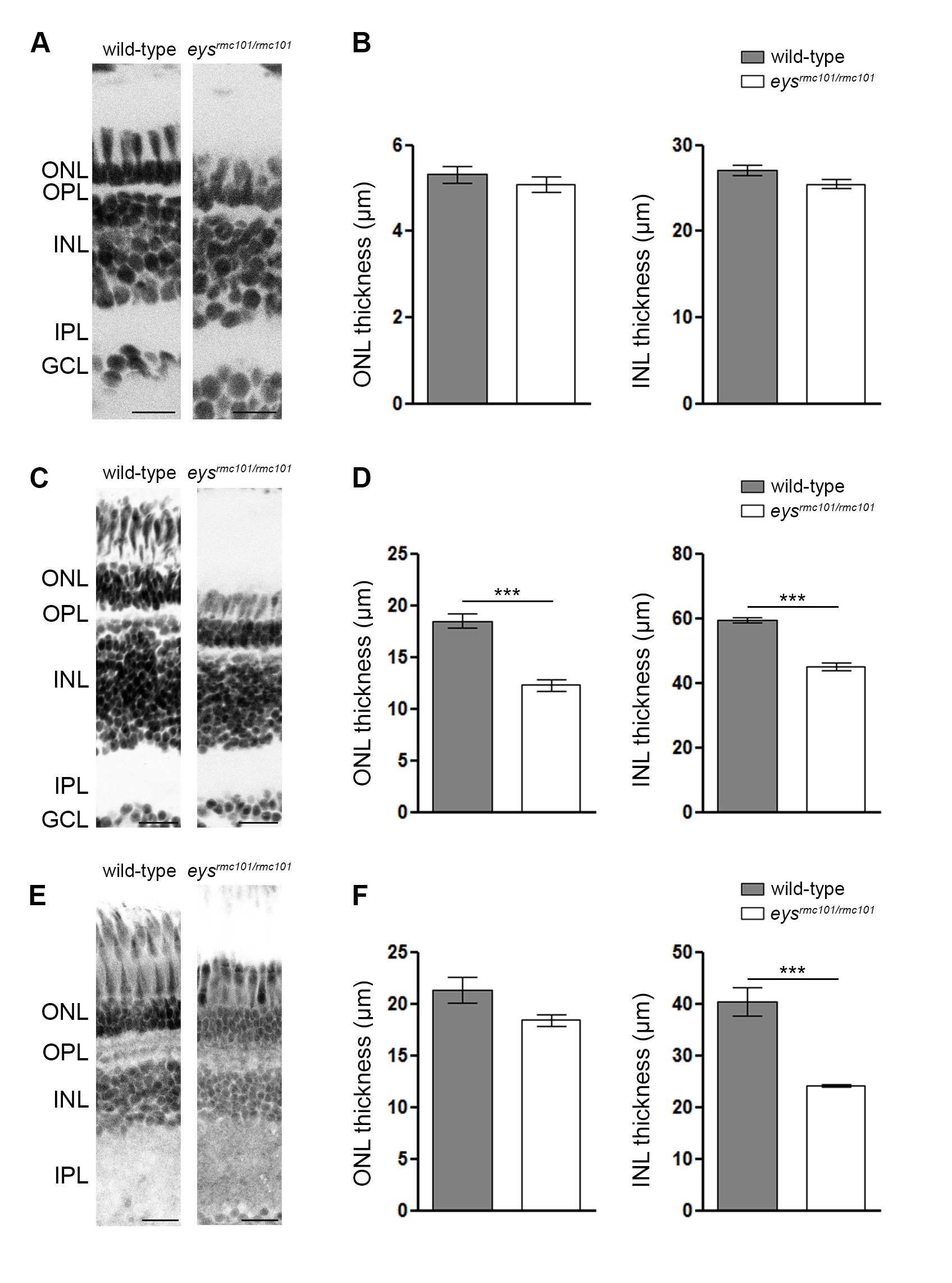
Fig. S3
Measurement of the thickness of outer and inner nuclear layers in eysrmc101/rmc101 and wild-type zebrafish.
(A, C, E) Representative images of wild-type and eysrmc101/rmc101 zebrafish retinas at (A) 5 dpf, (C) 2 mpf, and (E) 5 mpf. Nuclear layers were stained with DAPI and inverted to grey images. ONL: outer nuclear layer; OPL: outer plexiform layer; INL: inner nuclear layer; IPL: inner plexiform layer; GCL: ganglion cell layer. Scale bars (A): 10 μm. Scale bars (C, E): 20 μm. (B, D, F) Measurements of ONL and INL thickness in wild-type and eysrmc101/rmc101 zebrafish at (A) 5 dpf, (C) 2 mpf, and (E) 5 mpf. Asterisks indicate statistical significance (*** = p<0.0001) using Mann-Whitney U test.