
|
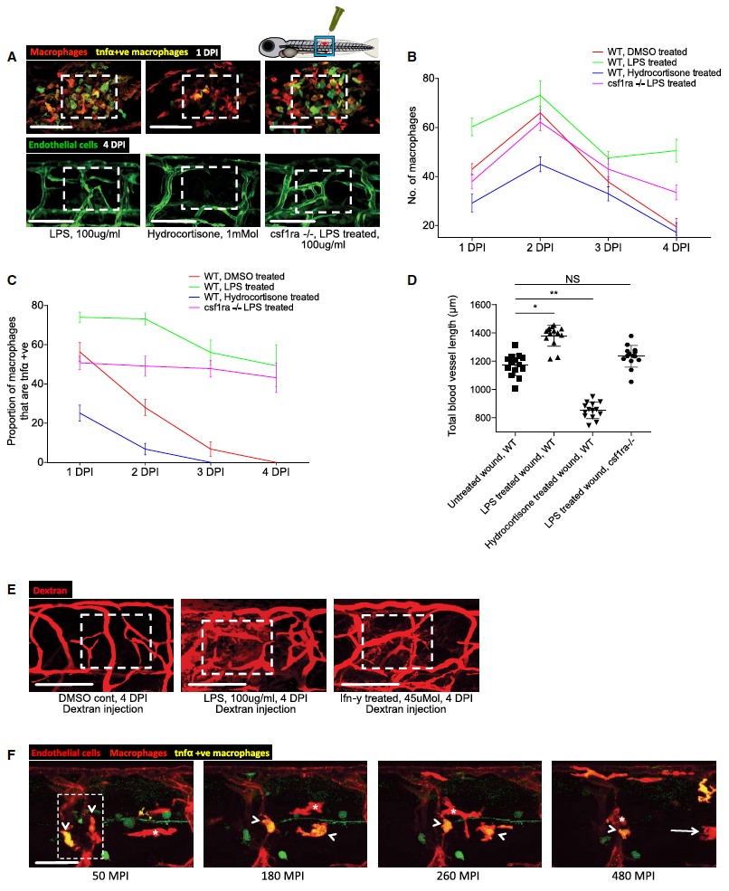
Fig. S4
Manipulation of normally dynamic macrophage phenotype towards a pro-inflammatory state alters the extent of wound angiogenesis and the patency of vessels.
A Representative confocal projection images taken from either 1 DPI Tg(mpeg:mCherry); Tg(tnfa:GFP) transgenic zebrafish (top panel) or 4 DPI Tg(fli:GFP) transgenic fish (bottom panel), wild type or csf1ra/ mutant, treated as indicated with LPS or hydrocortisone. Boxed area denotes wound site. B Quantification of number of macrophages at the wound site from 1 to 4 DPI, measured from images represented in (A) and Fig 4. N = 14 independent fish per timepoint per condition. C Quantification of proportion of macrophages that are tnfa positive at the wound site from 1 to 4 DPI, measured from images represented in (A) and Fig 4. N = 14 independent fish per timepoint per condition. D Quantification of total blood vessel length at 4 DPI, measured using Angioanalyser from images represented in (A). N = 14 independent fish per timepoint per condition. Statistical significance, as determined by one-way ANOVA, is P ≤ 0.0001. Subsequent Bonferroni multiple comparison test, determines level of significance, as indicated. Significance values: *P ≤ 0.05, **P ≤ 0.001. E Representative confocal projection images of 30-gauge needle wounded Tg(fli:GFP) zebrafish larvae, treated with DMSO, LPS or Ifn-c from moment of injury, loaded with dextran (red) one hour prior to imaging, imaged at 4 DPI. Boxed area indicates wound site. F Representative confocal projection images taken from laser wounded Tg(kdrl:mCherry-CAAX); Tg(mpeg:mCherry); Tg(tnfa:GFP) transgenic zebrafish, imaged 30– 480 MPI. Similar early recruitment was observed with respect to tnfa-positive macrophages (arrowheads) versus tnfa-negative macrophages (asterisks), complementing our results from Movie EV8. As time progresses, some tnfa-positive macrophages diminish their GFP expression as they leave the site of injury (arrow). Boxed area denotes wound site. N = 5 independent fish.