
|
Fig. S2
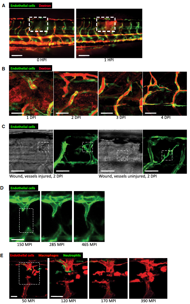
Dynamic vessel sprouting in response to tissue damage results in re-establishment of vessel patency within four days in zebrafish wounds.
A Representative fluorescent stereomicroscope images taken from 30-gauge needle-stick wounded Tg(fli:GFP) zebrafish. High molecular weight dextran was loaded one hour prior to injury, with images taken immediately after and one hour after injury, showing leakage of dextran (red) into the wound site upon vessel damage. N = 12 independent fish. B Confocal projection images of a representative 30-gauge needle wounded Tg(fli:GFP) zebrafish, loaded with dextran one hour prior to imaging, showing the typical time course of blood vessel leakiness over 4 DPI following needle-stick injury as per (A). Vessels typically cease leaking dextran by 4 DPI. N = 12 independent fish per timepoint. C Confocal projection images of a representative fine tungsten needle wounded Tg(fli:GFP) zebrafish, with damage induced to an existing vessel or between existing vessels. DIC images are provided as a guide to injury positioning. N = 8 independent fish per condition. D Representative confocal projection images taken from timelapse movies of a laser injured Tg(fli:GFP); Tg(mpx:GFP) zebrafish, 4 DPF, imaged at 150–465 MPI, showing the dynamic nature of vessel tip extensions. N = 10 independent fish. E Representative confocal projection images taken from timelapse movies of a laser injured Tg(kdrl:mCherry-CAAX); Tg(mpx:GFP); Tg(mpeg:mCherry) transgenic zebrafish, 4 DPF, imaged at 30–420 MPI (Movie EV5), to complement our dynamic studies of neutrophils and macrophages observed in Movie EV2. N = 5 independent fish.