
|
Fig. 6
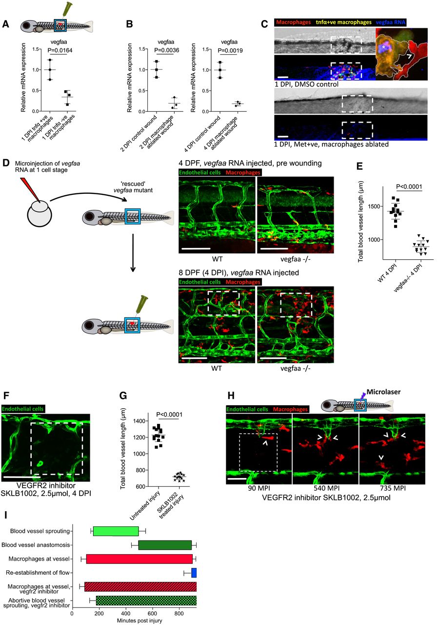
Zebrafish wound angiogenesis is driven by wound inflammatory macrophage‐mediated vegf signalling
Gene expression for FAC‐sorted zebrafish wound macrophages from Tg(tnfα:GFP); Tg(mpeg:mCherry) zebrafish, extracted at 1 DPI following needle‐stick injury. tnfα‐positive inflammatory macrophages express higher levels of vegfaa than tnfα‐negative non‐inflammatory macrophages. N = 50 independent fish per condition, 3 replicates. Statistical significance is indicated, as determined by two‐tailed t‐test.
Gene expression for whole‐dissected zebrafish needle‐stick wounds from Tg(mpeg:KalTA4); Tg(UAS:nfsB‐mCherry) zebrafish following treatment with metronidazole, versus control DMSO‐treated wounded fish, at 2 DPI and 4 DPI. Macrophage absence from wounds results in an overall decrease in vegfaa levels at the wound site. N = 30 independent fish per condition, three replicates. Statistical significance is indicated, as determined by two‐tailed t‐test.
Representative confocal projection images taken from needle‐stick wounded Tg(mpeg:KalTA4); Tg(UAS:nfsB‐mCherry); Tg(tnfα:GFP) triple transgenic zebrafish, DMSO control or metronidazole treated indicated, upon which vegfaa WISH was performed. Inset image shows high levels of vegfaa expression (blue) in tnfα‐positive macrophages (yellow, asterisk) versus low levels in tnfα‐negative macrophages (red, arrowhead). Boxed area denotes wound site. N = 12 independent fish per condition.
Schematic and representative confocal images showing rescue experiment and subsequent needle‐stick injury performed on vegfaa homozygous mutant zebrafish or siblings, transgenic for Tg(etv2:GFP). Embryos derived from an incross of vegfaa+/− adults were injected with “rescue” mRNA encoding for vegfaa‐165 at the one‐cell stage. At 4 DPF, these mutant fish had an impaired but functional complement of ISVs. Needle‐stick injuries performed on these mutants and their siblings at 4 DPF indicated a failure of wound angiogenesis by 4 DPI, despite a relatively normal inflammatory response. Boxed area denotes wound site.
Quantification of total blood vessel length at 4 DPI, measured from images represented in (D), using Angioanalyser. N = 12 independent fish per condition. Statistical significance is indicated, as determined by two‐tailed t‐test.
Representative confocal projection image taken from needle‐stick wounded Tg(fli:GFP) transgenic zebrafish, imaged 4 DPI and treated with vegfr2 inhibitor SKLB1002 from the moment of injury. Boxed area denotes wound site.
Quantification of total blood vessel length, measured from images represented in (F) using Angioanalyser. N = 14 independent fish. Statistical significance is indicated, as determined by two‐tailed t‐test.
Representative confocal projection images taken from laser wounded Tg(fli:GFP), Tg(mpeg:mCherry) double‐transgenic zebrafish, imaged 30–930 MPI and treated with SKLB1002 from moment of injury. Macrophages still associate with damaged vessels (arrowheads), but these vessels do not sprout appropriately and fail to repair. Boxed area denotes wound site.
Graphical representation of common cellular interaction events and their time course, measured from movies represented in (H) and compared to events observed in Fig 3C. N = 8 independent fish.