
|
Fig. 4
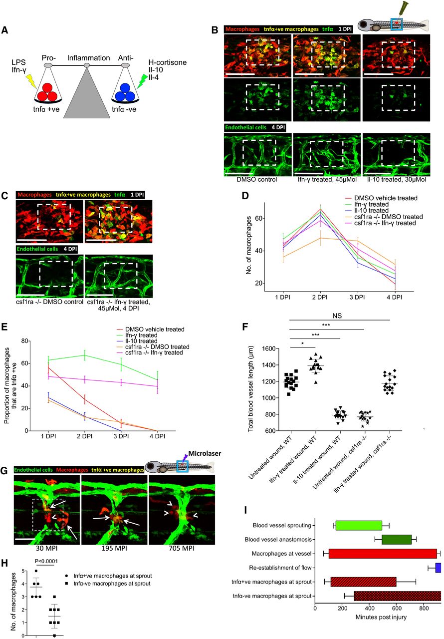
Manipulation of wound inflammation and macrophage phenotypic state affects the extent of wound angiogenesis
Schematic showing the factors used to skew macrophage phenotype towards either pro‐inflammatory or anti‐inflammatory states.
Representative confocal projection images of needle‐stick injured zebrafish, taken from either 1 DPI Tg(mpeg:mCherry); Tg(tnfα:GFP) transgenic zebrafish (top panel) or 4 DPI Tg(fli:GFP) transgenic zebrafish (bottom panel), treated, as indicated, with DMSO (control), Ifn‐γ or Il‐10. Boxed area denotes wound site.
Representative confocal projection images of needle‐stick injured zebrafish taken from either 1 DPI Tg(mpeg:mCherry); Tg(tnfα:GFP) transgenic fish (top panel; middle panel for GFP only) or 4 DPI Tg(fli:GFP) transgenic fish (bottom panel), csf1ra−/− mutant and treated with Ifn‐γ. Boxed area denotes wound site.
Quantification of number of macrophages at the wound site between 1 and 4 DPI, measured from images represented in (B and C). N = 14 (from B) and N = 16 (from C) independent fish per timepoint per condition.
Quantification of proportion of macrophages that are tnfα positive at the wound site from 1 to 4 DPI, measured from images represented in (B and C). N = 14 (from B) and N = 16 (from C) independent fish per timepoint per condition.
Quantification of total blood vessel length at 4 DPI, measured from images represented in (B and C), using Angioanalyser. N = 14 (from B) and N = 16 (from C) independent fish per timepoint per condition. Statistical significance, as determined by one‐way ANOVA, is P ≤ 0.0001. Subsequent Bonferonni multiple comparison test, determines level of significance, as indicated. Significance values: *P ≤ 0.05, ***P ≤ 0.0001.
Representative confocal projection images taken from laser wounded Tg(fli:GFP); Tg(mpeg:mCherry); Tg(tnfα:GFP) triple transgenic zebrafish, imaged 30‐930 MPI. Boxed area denotes wound site. tnfα‐positive, inflammatory macrophages (arrows) associate with damaged, sprouting blood vessels earlier and in larger numbers than tnfα‐negative, non‐inflammatory macrophages (arrowheads).
Quantification of numbers of inflammatory versus non‐inflammatory macrophages associated with damaged, sprouting blood vessels, measured from images represented in (G). N = 8 independent fish. Statistical significance is indicated, as determined by two‐tailed t‐test.
Graphical representation of common cellular interaction events and their time course, measured from movies represented in (G) and compared to events observed in Fig 3C. N = 8 independent fish.