
|
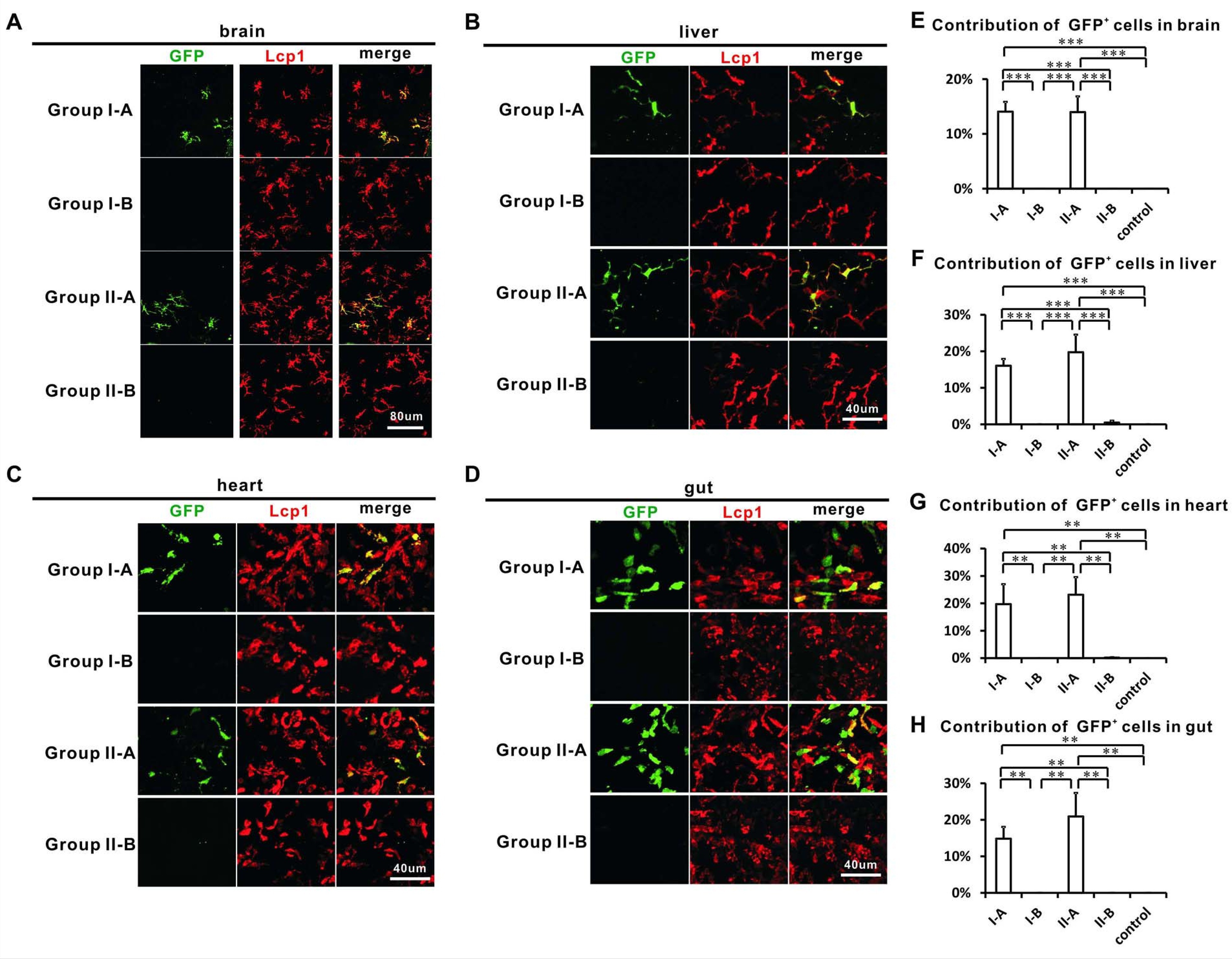
Fig. 5
Adult tissue-resident macrophages in the brain, liver, heart and gut correlate with HSCs.
(A–D) Confocal images show that GFP+ cells are found in the brain (A), liver (B), heart (C) and gut (D) of Group I-A and II-A, but not I-B and II-B. Most GFP+ cells are Lcp1+ with the exception of gut GFP+ cells which only partially overlap with Lcp1 signals. (E–H) Quantification of the contribution of GFP+ cells in all Lcp1 positive leukocytes in the brain (E), liver (F), heart (G), and gut (H) of adult Group I-A, I-B, II-A, II-B, and non-heat-shocked control group (n = 4 for each group of brain and liver; n = 3 for each group of heart and gut). Error bars represent mean SEM. ***p<0.001; **p<0.01.