
|
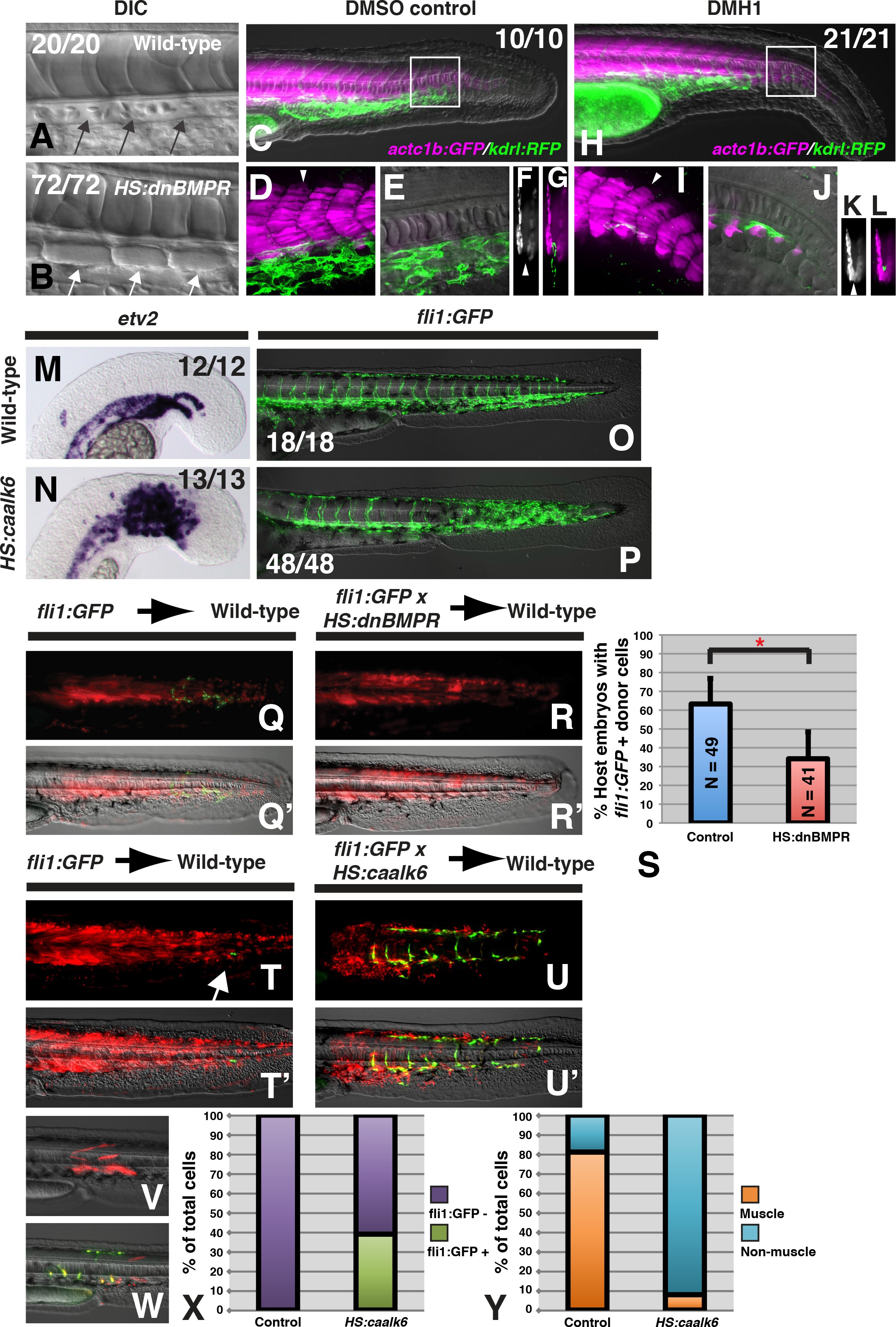
Fig. 1
MBMP signaling is necessary and sufficient for endothelial fate specification in tailbud-derived mesoderm.
(A) Wild-type sibling embryos heat-shocked at the 12-somite stage exhibit normal formation of the dorsal aorta (black arrows, 20/20 normal). (B) HS:dnbmpr embryos heat-shocked at the 12-somite stage have ectopic segmented somite tissue where the dorsal aorta normally forms (white arrows, 72/72 with ectopic somite tissue). (C–L) Loss of BMP signaling using the small molecule DMH1 phenocopies HS:dnbmpr embryos. Embryos transgenic for both the actc1b:GFP (muscle, magenta) and kdrl:rfp (endothelium, green) transgenes were treated with DMSO (C–G) or DMH1 (H–L). A confocal Z-projection of the boxed region in C shows the presence of both muscle and endothelium in control DMSO treatment. A single z-slice at the midline shows the presence of endothelium and absence of muscle, which can also be observed in a digital cross section at the level of the white arrowhead in panel D. A confocal z-projection of the boxed region in H shows the presence of muscle and large reduction in endothelium (I). A single z-section at the midline shows the reduction of endothelium is accompanied by ectopic midline muscle formation, also observed in the digital cross-section at the level of the white arrowhead in panel I. (M, N) Transgenic HS:caalk6 embryos heat-shocked at the 12-somite stage exhibit expansion of the endothelial marker etv2 into the pre-somitic mesoderm 5 hr after the heat-shock (control N = 12, HS:caalk6 N = 13). (O, P) At 36 hpf, HS:caalk6 embryos heat-shocked at 12-somite stage have a dramatic expansion of fli1:GFP expression in posterior regions that would normally form somites, whereas there is no effect on anterior somites that formed before the heat-shock (Control N = 18, HS:caalk6 N = 48). (Q–R’) Rhodamine dextran (red) labeled fli1:GFP donor cells were transplanted into unlabeled wild-type host embryos to monitor for contribution of transplanted cells to endothelium. (Q, Q’, S) Control cells contribute to endothelium in 63% of host embryos (N = 49). (R, R’, S) Heat-shock induction of dnbmpr at the 12-somite stage significantly (p=0.0107) reduces the percentage of host embryos (34%) that have donor-derived endothelium (N = 41). (T–U’) Induction of endothelium by BMP signaling is cell-autonomous, as exhibited in HS:caalk6 x fli1:GFP cells transplanted wild-type host embryos. Host embryos were heat-shocked at the 12-somite stage and assayed for fli1:GFP expression at 36 hpf. (U, U’) HS:caalk6 transgenic cells do not contribute to somites and instead give rise to endothelium. One-cell transplants were done to quantify fate changes after BMP activation (W) compared to controls (V). 12-somite stage BMP activation resulted in 39% fli:GFP positive cells (four embryos, 49 cells), compared to 0% in control transplants (three embryos, 36 cells, p<0.0001) (X). The fate of control transplanted cells was 81% muscle, whereas only 8% of HS:caalk6 cells adopted a muscle fate (p<0.0001) (Y). All embryos are pictured from a lateral view with the head to the left, except for F, G, K, and L which are digital transverse sections.