
|
Fig. 1
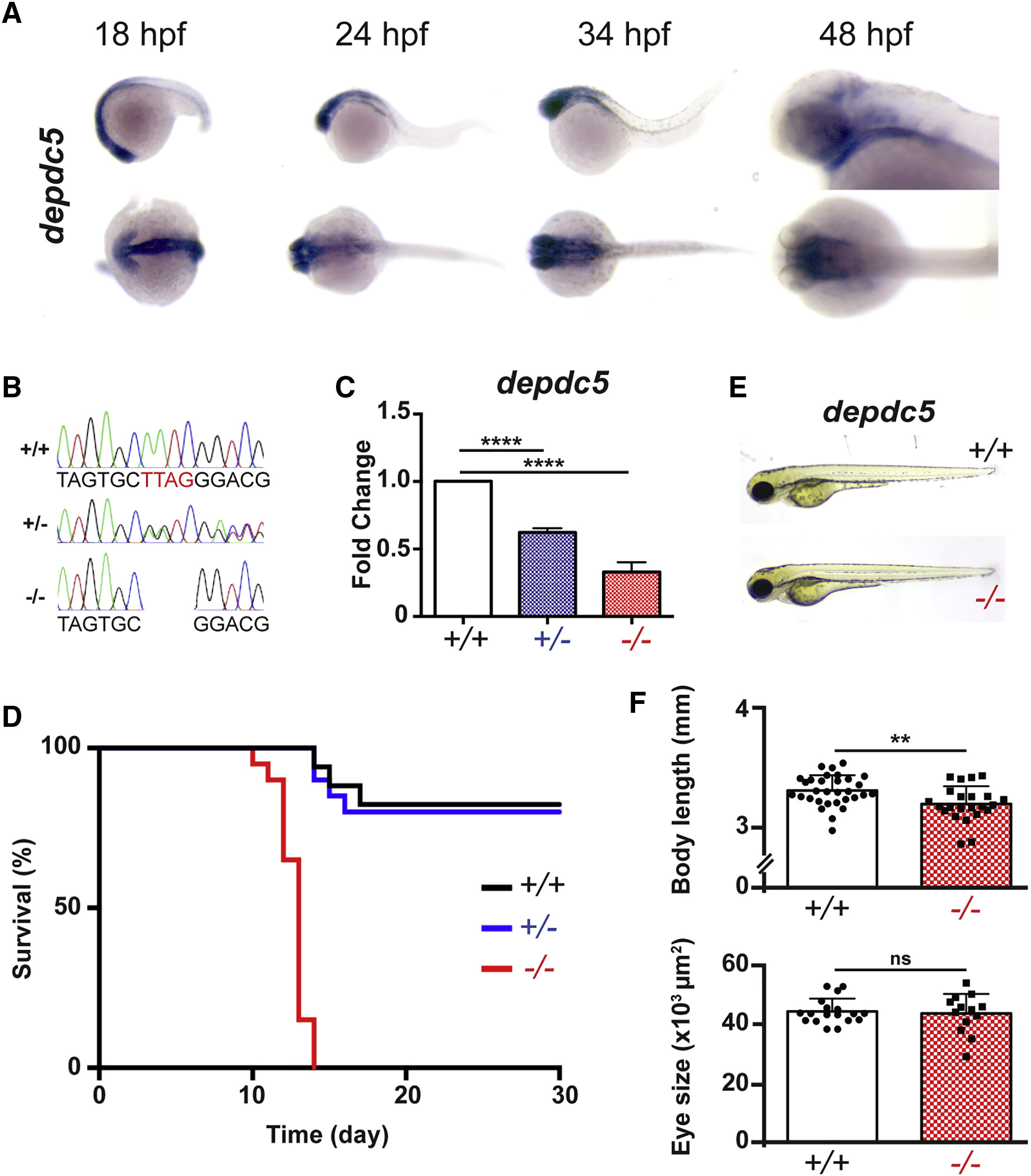
depdc5 Is Essential for Survival
(A) Whole-mount in situ hybridization showing expression of depdc5 at different stages of development.
(B) Electropherograms showing confirmation of the 4-bp insertion by Sanger sequencing.
(C) Expression of depdc5 transcript in 7-dpf depdc5+/+, +/−, and −/− larvae. N = 3; Student’s t test: p(WT versus heterozygote [HT]) = 0.0026; p(WT versus homozygote [HM]) = 0.001.
(D) Survival curve showing that depdc5−/− embryos have reduced survival, with most larvae dying around 14 dpf (N = 2, n = 15 per genotype).
(E) Images showing that depdc5−/− larvae do not show gross morphological changes at 3 dpf.
(F) Measurement of body length (top; n = 20/genotype; p = 0.0045 by unpaired Student’s t test) and eye size (bottom; n = 12/genotype; p = 0.7219 by unpaired Student’s t test) showed no significant change in either parameter.