
Image
|
Figure Caption
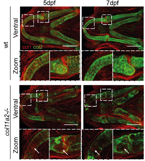
Fig. S4
Type 1 collagen is not increased in col111a2 mutants. Immunostaining for Type 1 (red) and Type II (green) collagen in wt and col11a2 -/- at 5 and 7dpf. Dashed insets show areas of higher magnification. White arrows indicate areas of Type II collagen loss, where Type I collagen is unchanged. Scale bar = 100 µm
Figure Data
Acknowledgments
This image is the copyrighted work of the attributed author or publisher, and
ZFIN has permission only to display this image to its users.
Additional permissions should be obtained from the applicable author or publisher of the image.
Full text @ Phil. Trans. Roy. Soc. Lond., Series B