
Image
|
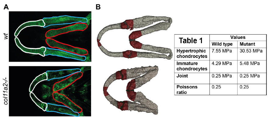
Figure Caption
Fig. S5
Segmentation of lower jaw elements and assignment of material property values. A) The Meckel's cartilage (white), palatoquadrate (blue) and ceratohyal (red) were segented from confocal images of 7dpf wt and col11a2 -/- zebrafish. B) Material properties from Atomic Force Microscopy were applied to the corresponding regions of the lower jaw. Dark red = immature chondrocytes in the joint regions, grey = hypertrophic chondrocytes towards the middle of the cartilage element, and green = joint. TAble 1) Material property values used for Finite Element model generation (MPa = megapascals).
Acknowledgments
This image is the copyrighted work of the attributed author or publisher, and
ZFIN has permission only to display this image to its users.
Additional permissions should be obtained from the applicable author or publisher of the image.
Full text @ Phil. Trans. Roy. Soc. Lond., Series B