
|
Fig. 4
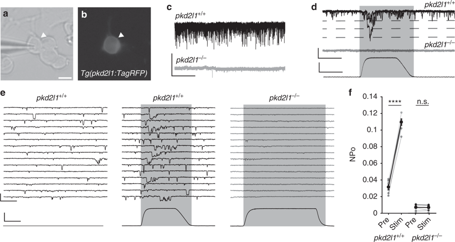
CSF-contacting neurons require Pkd2l1 to respond to mechanical pressure. a Transmitted light and b fluorescent images of a cultured CSF-cN from a Tg(pkd2l1:TagRFP) embryo. Scale: 5 µm. c Gap-free VC recordings from CSF-cNs in vitro show extensive single channel opening in pkd2l1+/+ but not pkd2l1−/− embryos, comparable to results in vivo. Scale: 2 s, 20 pA. d Gap-free VC recording from a cultured CSF-cN while a mechanical stimulus is applied, showing an increase in channel opening during the stimulus in pkd2l1+/+. Scale: top: 20 ms, 25 pA, bottom: 20 ms, 2 µm. e Gap-free recording from cultured CSF-cNs while a mechanical stimulus is applied. Scale: top: 20 ms, 25 pA, bottom: 20 ms, 2 µm. f Quantification of channel opening probability in response to mechanical stimulation of pkd2l1+/+ and pkd2l1−/− CSF-cNs (n = 7 pkd2l1+/+ CSF-cNs, pre vs. stim p = 1.6 × 10⁻⁶; n = 10 pkd2l1−/− CSF-cNs, pre vs. stim p = 0.76, paired t test). Error bars represent s.e.m.