
Image
|
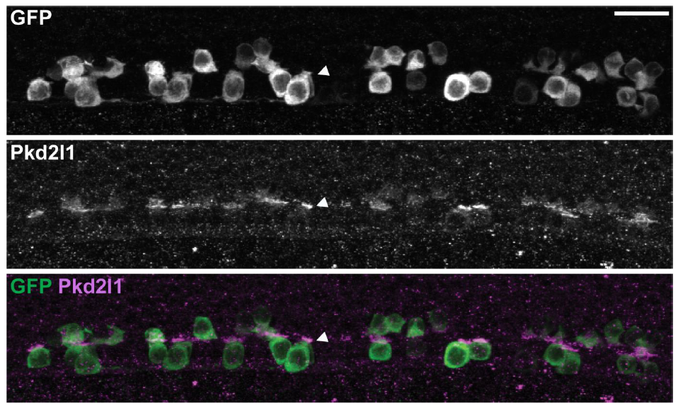
Figure Caption
Fig. S5
Pkd2l1 is correctly localized to the apical extension in the cfap298m304/tm304 mutant.
Pkd2l1 immunohistochemistry in Tg(pkd2l1:GCaMP5G; cfap298tm304/tm304) at 30 hpf. Top: GFP staining, middle: Pkd2l1 staining, bottom: merge. Arrowhead shows dense Pkd2l1 localization in the apical extension contacting the CSF in the central canal. Scale: 20 μm.
Acknowledgments
This image is the copyrighted work of the attributed author or publisher, and
ZFIN has permission only to display this image to its users.
Additional permissions should be obtained from the applicable author or publisher of the image.
Full text @ Nat. Commun.