
|
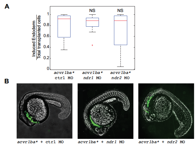
Fig. S7
Ndr1 and Ndr2 act redundantly to support the ability of acvr1ba* cells to internalize.
(A) Boxplot quantification of endoderm contribution of transplanted cells at 20 hpf. Data is shown as mean ± SEM of independent transplantation experiments with 16 embryos per condition. Student’s t-test was performed. NS, not significant.
(B) Representative image showing distribution of transplanted cells depicted in (A) at 18 hpf. acvr1ba*-expressing cells localized to the endoderm-derived tissue (green)in all three conditions, in contrast to the block of internalization when both Ndr1 and Ndr2 MO are combined in acvr1ba* cells (Fig. 2H). Lateral view, anterior to the left.