
|
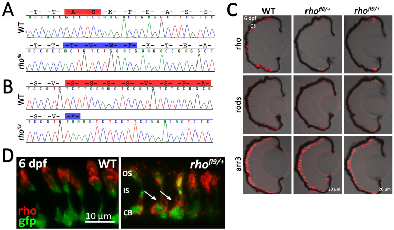
Fig. 4
Alteration of conserved C-terminal domains in rhofl8 and rhofl9. A, B: Chromatograms overlaid with amino acid sequences of 3′ rh1-1 in WT, the rhofl8 allele, and the rhofl9 allele. Predicted amino acid deletions are highlighted in red on the WT sequences; insertions are highlighted in blue on the mutant sequence. C: Confocal images of retinal sections of 6 dpf WT or heterozygous rhofl8/+ or rhofl9/+ larvae labeled with antibodies to Rho (1D1, red), rods (4C12, red), or Arr3a (Zpr-1, red) overlaid with bright-field microscopy. D: High magnification confocal images of 6 dpf WT or heterozygous rhofl9/+ larvae showing the rod-specific expression of EGFP (gfp, green) and immunolabeling for Rho (1D1, red). In WT retinas, the Rho immunolabeling is localized to the outer segment (OS), while in the mutant, Rho immunolabeling is localized to the inner segment (IS) and the cell body (CB; arrows).