
|
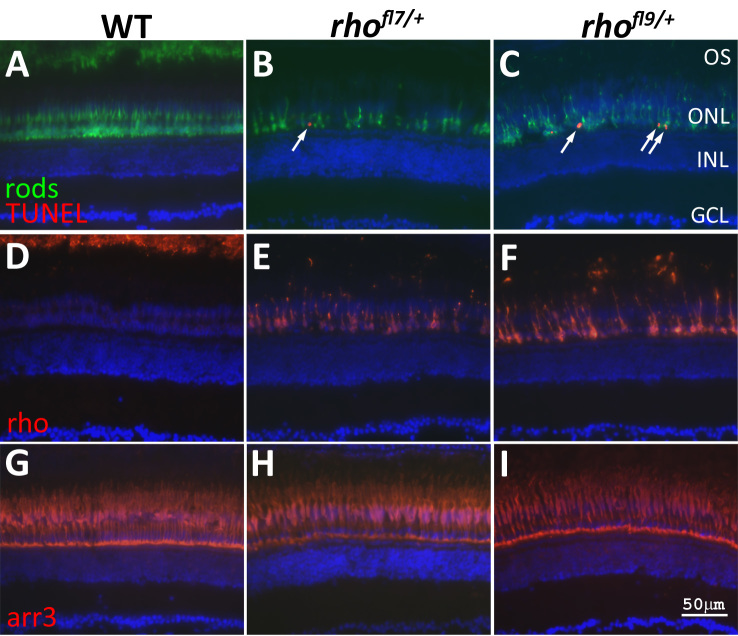
Fig. 5
rhofl7 and rhofl9 lead to increased TUNEL in adults. Retinal sections of WT (A,D,G) or heterozygous rhofl7/+ (B,E,H) and rhofl9/+ (C,F,I) adult zebrafish immunolabeled with antibodies to rods (4C12, green, A-C), Rho (1D1, red, D-F), or red/green cones (Arr3a/Zpr-1, red, G-I) and stained with TUNEL (red, A-C). All sections were counterstained with DAPI (blue). Immunolabeling for rods reveals fewer, less regularly arranged cells in the mutants compared to the WT. TUNEL-positive nuclei (arrows), positioned along the proximal region of the ONL, are only observed in the mutant retinas (A-C). In WT retinas, Rho immunolabeling is restricted to the ROS at the top of the panel D. Immunolabeling in rhofl7/+ and rhofl9/+ adults is localized to the cell bodies, and no outer segments are evident (E-F). Immunolabeling for red and green cones was indistinguishable across the samples (G-H). Abbreviations: ganglion cell layer, GCL; inner nuclear layer, INL; outer nuclear layer, ONL.