
|
Fig. S4
Further analysis of ATG MO hematopoietic phenotype.
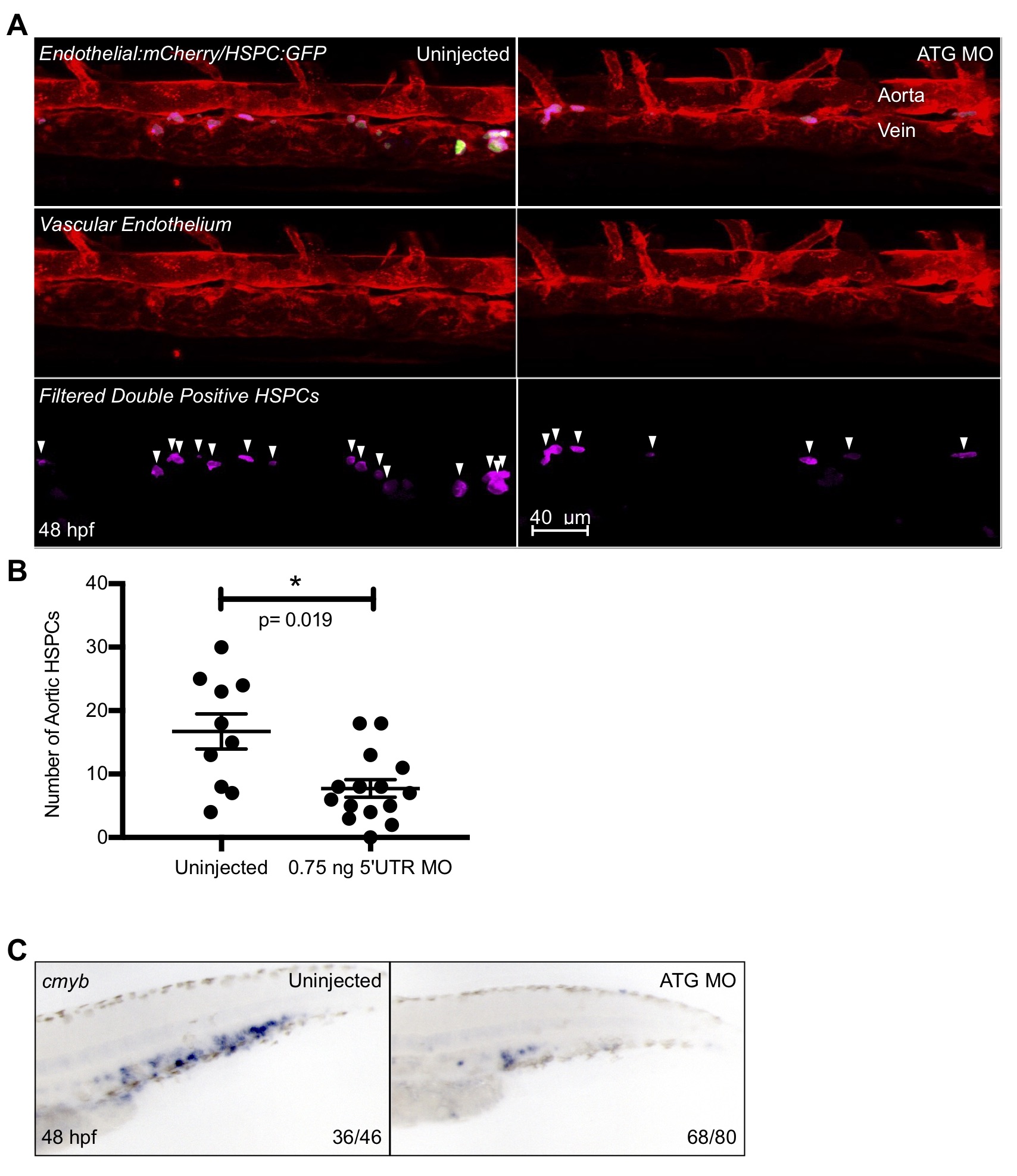
ATG MO embryos were subjected to WISH for the hematopoietic marker cmyb at 48 hpf (A). The caudal hematopoietic tissue of morphant embryos showed a distinct reduction of cmyb staining as compared to their uninjected siblings. The morpholino was also injected into Tg(CD41:GFP/kdrl:mCherry) embryos and double positive fish were imaged via confocal microscopy at 48 hpf and Imaris imaging software was used to remove GFP signal outside of the vasculature (B). The surfaces feature of Imaris was utilized to quantify double positive cells (shown here in pink), and the resulting data was graphed and statistically analyzed by a non-parametric t-test on Prism (C). Error bars are SEM. There was a small, but significant decrease in the number of HSPCs in the ATG morphant fish. Numbers in the lower right-hand corner of each image depict the number of embryos with the phenotype pictured out of the total number of embryos assayed in each condition.