
|
Fig. S1
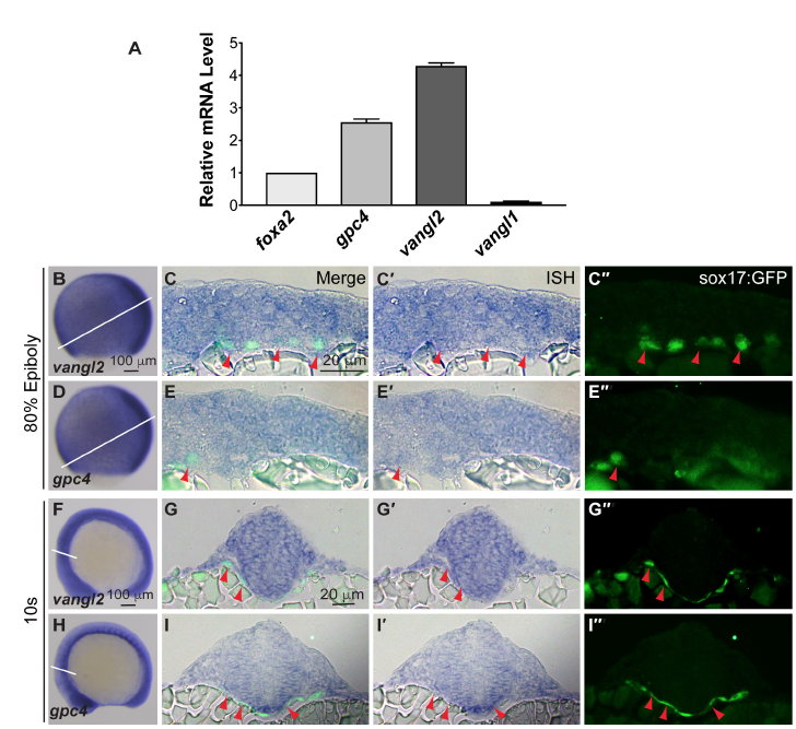
The expression of gpc4 and vangl2 during gastrulation and early segmentation.
(A) Expression of gpc4, vangl2 and vangl1 relative to that of foxa2, and endoderm marker, as determined by qRT-PCR, in GFP+ cells sorted from Tg(sox17:EGFP) embryos at 18s. Bars represent the mean±s.e.m. (B-I′′) The expression of gpc4 and vangl2 transcripts in Tg(sox17:EGFP) embryos at 80%E and 10 s, as detected by WISH. (B, D, F, H) Images of the whole embryo. White lines indicate the cross-sectional plane. (C-C′′, E-E′′, G-G′′, I-I′′) Transverse sections of the embryos. (C, E, G, I) Overlays of anti-GFP immunofluorescence staining (sox17:EGFP panels) and ISH for vangl2 and gpc4 (ISH panels), in endodermal cells (red arrowheads).