
|
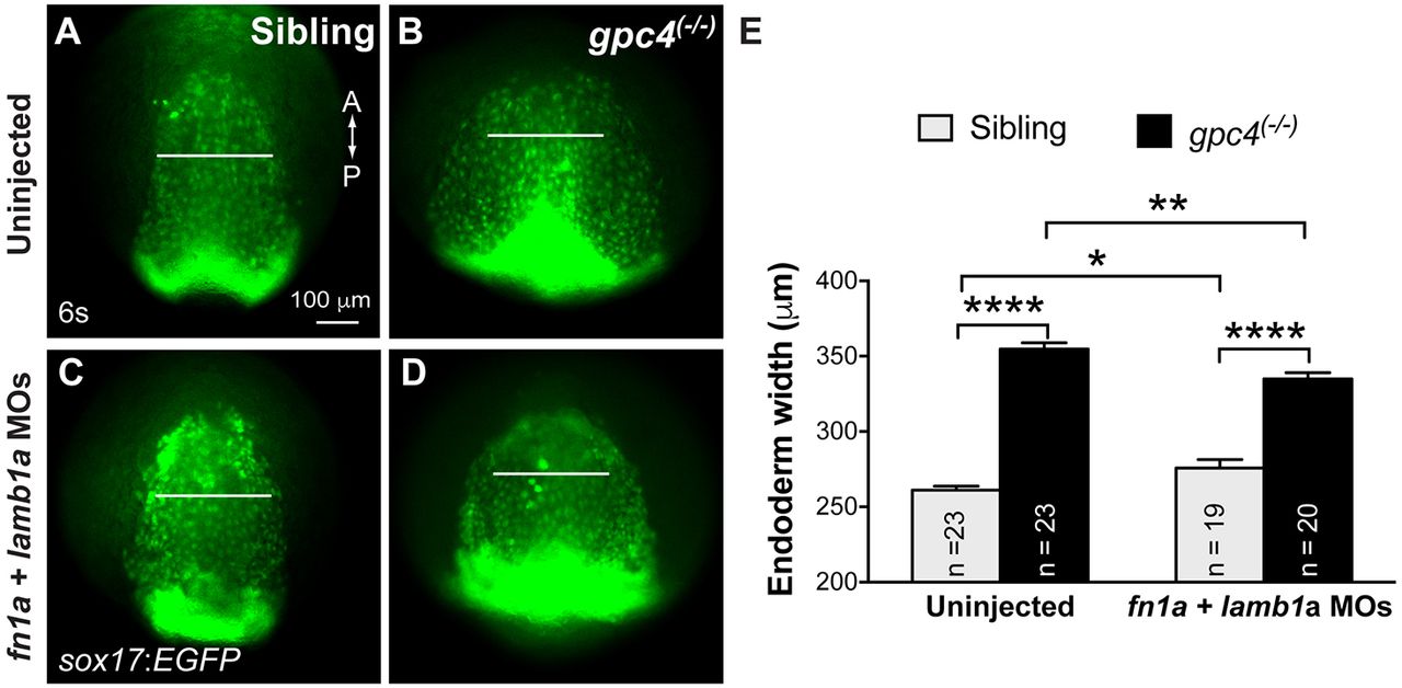
Fig. 6
Endodermal defects in gpc4 mutants are suppressed by knocking down fn1a and lamb1a. (A-D) Epifluorescence still images of the anterior region of the endodermal sheet in 6s embryos derived from crossing of gpc4/Tg(sox17:EGFP) heterozygous fish injected with or without MOs targeting fn1a and lamb1a (5 ng each). Anterior-dorsal view. White lines of equivalent length indicate width of the anterior endodermal sheets. A, anterior; P, posterior. (E) Average width of anterior endodermal sheet in embryos shown in A-D. Number of embryos analyzed in each group is indicated. *P<0.05, **P<0.01, ****P<0.0001; Student's t-test.